13. Progress in SSRL Operations
by Piero Pianetta
Appendix B Self-Evaluation FY2006
Return to Table of
Contents
FY2006 User Experimental Run
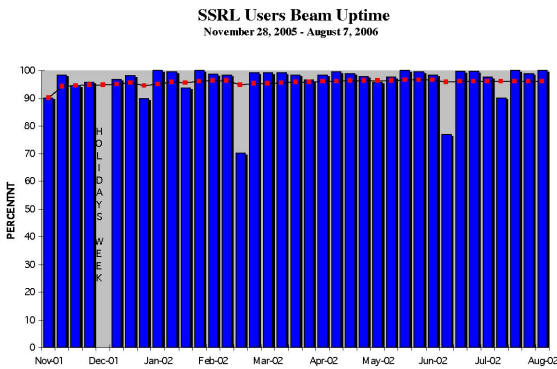
In the FY2006 user run, the facility proved to be
exceptionally reliable, providing very stable beam for a
very high fraction (96.2%) of the scheduled time. This
is an exceptional achievement for a new storage ring.
The user run commenced on November 28, 2005 and
continued through August 7, 2006, and the SPEAR3 storage
ring operated at 3 GeV/100 mA and provided 60+ hour life
times. (The average uptime over the past five years was
96%.) During the FY2006 run, scientists on 345 different
proposals received beam time in a total of 1,002
experimental starts involving approximately 1,700 users,
with approximately 900 users on-site or remote accessing
beam line equipment. Approximately 66% of the users came
from universities and other laboratories in the United
States, 15% from DOE and US government laboratories, 5%
from US industry, and 14% from international
institutions.
Distribution of Proposals Receiving Beam:
Materials Science 14%
Physics 5%
Chemistry 16%
Polymers
2%
Medical Applications 5%
Biology/Life Sciences 45%
Earth Sciences 3%
Environmental Sciences 5%
Optics 1%
Engineering 3%
Other 1%
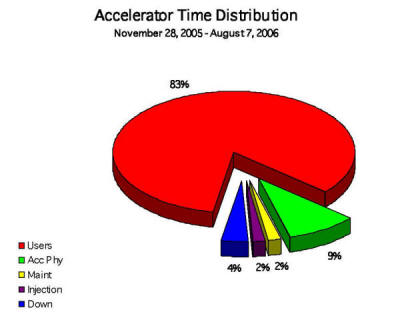
SPEAR3 Accelerator Improvements
The accelerator improvements for SPEAR3 are focused in
four areas: 1) characterizing and improving accelerator
performance and reliability at 100 mA to maximize beam
quality for users; 2) developing and implementing a new
lattice optics configuration that accommodates a
magnetic chicane having two small vertical beam size
waists (the double-waist chicane optics, or DWC optics)
in the east long straight section; 3) characterizing and
optimizing accelerator operation at 500 mA; and 4)
preparing for the implementation of top-off injection
with beam line stoppers open.
Injector –
Work continued in FY2006 to improve the reliability and
operation of the injector at 3 GeV. The injection timing
system was modified to equalize booster ramping
efficiency for all bucket timings. Work continued to
characterize and stabilize gun and linac operation. A
feedback system was implemented to stabilize the booster
White Circuit. Booster performance was assessed and
plans for its realignment were made. A study of the
stability of all injector power supply systems was
initiated. Additional improvements include rebuilding
the BTS injection line (to remove several vacuum
windows), upgrading transport line beam position and
intensity monitors, enhancing the stability and
monitoring of powered devices, completing a pulsed
signal monitoring system, implementing an EPICS control
system access to injector component control and readback
variables, and installing a second klystron for the
gun/linac system. The design of simpler and more robust
booster kicker pulsers has begun and plans for
developing a laser-heated-cathode rf gun are in
progress. The Accelerator Physics group is studying
booster beam capture efficiency and has proposed a minor
lattice modification that could improve performance.
Studies of other booster improvements, including the
powering of unused sextupole magnets and the possible
addition of a subharmonic rf cavity, are under way. The
injector improvement project will continue in FY2007 and
beyond.
Turn-Turn Beam Position Monitors (BPMs) –
Commissioning and development of the user software
interface for the turn-turn BPM system is continuing in
FY2006. The test tone calibration system will be
commissioned and the true performance of the processors
is being quantified. The turn-turn BPMs will then be
integrated into the orbit feedback system.
Synchrotron Light Monitor (SLM)
–
The SLM beam line received first light in January 2006.
The optical bench components were commissioned and the
beam line component alignment was refined. The first
measurement of beam bunch length was performed during
the user run and low alpha experiments were performed in
which the bunch length was shorted to 6.5 ps with 100 mA
stored current in SPEAR. The upgrade and realignment of
the BL2 pinhole camera system was completed in early
spring of 2006.
Beam Scrapers
–
The new horizontal and vertical beam scrapers were
commissioned and used to characterize beam lifetime,
injection apertures, and minimum apertures for future
insertion devices. The scrapers will continue to be a
valuable diagnostic for beam size, lifetime and aperture
studies.
LION Development –
Commissioning of the Long Ion Chamber (LION) system
continued during the FY2006 user run. Tests of the
system with high injected beam current were carried out
in early spring 2006. The LION system must be connected
to the Beam Containment System before the injected beam
current limit can be raised to enable faster 500-mA fill
times.
High-Current SPEAR3 Tests – SSRL received authorization from the DOE Site Office to
conduct SPEAR3 operation with currents up to 500 mA,
above the official safety envelope value of 100 mA
allowing routine running of SPEAR3 with beam lines
closed for accelerator studies. A successful test of the
Double Waist Chicane lattice (see below) at 500 mA was
conducted in February 2006 with no observed problems.
Orbit Control
– The first phase of the fast orbit feedback system,
using only the 54 Bergoz averaged-orbit BPMs, was
completed. A test of the system demonstrated successful
feedback operation with a bandwidth approaching 100 Hz.
Further work was done to optimize the SVD-derived
inverse response matrix and digital filters as well as
to maximize the frequency response
of the orbit corrector power supplies. Work continued in
order to provide an operator interface anddata
acquisition applications for the new fast orbit feedback
system.
The orbit monitor processing system has a temperature
dependence that has been demonstrated to cause orbit
instability when the feedback system is active. A
project to build temperature-controlled rooms and
install finer temperature regulation in the processor
equipment racks was completed in time for testing before
the end of the run. Initial tests showed that the
cooling system reduced the processor temperature
variation from several degrees Celsius to less than a
degree, thus decreasing the processor
temperature-related orbit instability. Further tests
during the 2007 run will quantify the degree of
improvement.
Hydrostatic level sensors (HLS) were installed a few
strategic areas in the SPEAR3 tunnel to monitor the
vertical motion of tunnel floor and some beam line
components. If this system proves to be useful for
detecting component motion, more sensors will be added
in the future and their signals will be used in the
orbit feedback system.
Double Waist Chicane (DWC) Lattice
– The DWC optics, temporarily installed without chicane
magnets during the FY2005 shutdown, was tested
successfully for the first time in December 2005. While
the lifetime was a few percent less than that for the
normal lattice, this was anticipated in the optics
studies and is acceptable. This lattice was
characterized and optimized so that it will be used with
the new BL12 undulator and beam line which was installed
during the 2006 summer shutdown. It has already been
demonstrated to work without problems at 500 mA and with
the “dispersion leak” optics variation that reduces the
emittance from 18 nm-rad to 12 nm-rad. The design and
fabrication of vacuum chamber and chicane magnets needed
for the full DWC implementation are being completed
during the 2006 shutdown. Most of the components have
been installed, together with the BL12 in-vacuum
undulator. The installation of the beam line that
utilize the downstream chicane straight section is
underway.
Top-Off Injection –
The design of the system needed for top-off injection
with beam line stoppers open was begun in FY2006. This
injection mode, with minimal interruption to users, will
enable more frequent beam injection to limit beam
current variation, minimizing the variation of thermal
power on beam line optical components and improving beam
stability for users. Work has begun to improve the
injector systems to accomplish this injection mode (see
above). An extensive study of beam loss modes was
initiated to determine what radiation safety components
will be required to inject beam into SPEAR3 with open
beam line radiation stoppers. Significant changes to the
Personnel Protection System, including the Septum
Interlock, will be needed to implement the top-off
system. This work will enable the first phase of top-off
injection - a mode that will maintain beam current
constancy in SPEAR3 to a few percent. A second phase of
top-off development that would enable maintaining SPEAR3
beam current constancy to less than 1% will most likely
require large-scale improvements to the electron gun and
possibly to the booster. These changes will not commence
until FY2007.
SPEAR3 Performance and Lattice Upgrades
– The accelerator physics and engineering groupscontinued to study and tune the SPEAR3 accelerator
to maximize its performance and stability. This work
includes investigating the sources of beam instability
(power supplies, rf systemcomponent vibration and
temperature-related motion, etc) and ongoing efforts to
characterize beam dynamical behavior as a function of
lattice and insertion device parameters. With regards to
the latter, a detailed study of the deleterious effects
of the new BL13 EPU on beam properties, and possible
cures, is in progress in collaboration with physicists
at other light sources. The results of this study will
be incorporated into the design of the BL13 EPU system.
An investigatory study of the possibility of circulating
short bunch electron bunches (<~1 ps) in the SPEAR3
storage ring was initiated. A new isochronous lattice
configuration was developed that would preserve the
short bunch length, and provide the option of
circulating the bunches for some small number of
revolutions (<1000), as opposed to storing the beam,
will be analyzed.
Gun Test Facility
– The GTF continued to support the LCLS injector group
through FY2006. Experiments performed included novel
in situ
methods to improve the cathode performance,
experiments to eliminate the correlated energy spread
produced by the gun, and beam-based screen resolution
measurements. New diagnostics will continue to be
developed including the electro-optic bunch length
measurement as well as testing new diagnostics such as
digital cameras and bunch charge monitors. The laser
will continue to be used to test transverse pulse
shaping techniques, the LCLS streak camera and methods
to improve the laser pointing stability.
Additional ultrafast electron diffraction experiments
were also planned. Improved detectors were tested and
the pulse length reduced to improve the temporal
resolution. The useful operating range of the gun for
electron diffraction has also been explored.
Beam Line and Facilities Improvements
BL1, 2, 3, 8 (bend beam lines)
–
Upgrade activities on bend magnet beam lines 1, 2, and 8
were limited to those essential to keep the beam lines
operating with higher SPEAR3 current. In particular,
during the 2006 shutdown, the BL2 beam position monitor
was upgraded for improved stability and the BL8 beam
position monitor shielding was upgraded for 500 mA
operations. BL3 will remain closed.
BL4
–
The BL4 upgrade continued with the fabrication of
components and is scheduled for installation during
FY2007. The BL4 upgrade is partially funded by DOE BER.
BL5 –
The BL5-1 M3
refocusing mirror system has been installed and
commissioned as has the shielding for 500 mA operation.
No other significant upgrades are scheduled.
BL7 –
The 500-mA upgrade installation was completed with the
temporary BL7-2 LN monochromator and the beam line was
commissioned. Assembly of the BL7-2 sagittal focusing
monochromator will be completed and the monochromator
will be installed and commissioned at the beginning of
the FY2007 run. BL7 and BL 7-3 upgrades are largely
funded by NIH NCRR.
BL10
– While no significant upgrades are planned, some
cooling enhancements of zero order beam masks downstream
of the BL10-1 monochromator have been scheduled.
New beam lines under development:
BL12
–
The in-vacuum ID was delivered and installed in the ring
as were the remaining dipole magnets and associated
vacuum chamber required to produce the SPEAR3 orbit
chicane. The hutches are being erected. The remaining
optical components will installed during October 2006.
It is anticipated that the beam line will start
commissioning by November 2006. The computing
infrastructure required to support this beam line is
being installed. A large-area CCD detector was procured
with an October 2006 delivery date. This beam line is
funded by the California Institute of Technology through
a gift from the Gordon and Betty Moore Foundation.
BL13
–
The ID fabrication will continue. The beam line front
end design will be completed and fabrication will
commence. The M0
mirror system will be designed and the optic ordered. Design of the in-alcove beam transport system will start.
Rather than order a new monochromator for BL13, it has
been decided to relocate the BL5-1/5-2 spherical grating
monochromator, associated slits, and refocusing optics
to BL13. This relocation is planned for the summer 2007
shutdown. During the remainder of FY2006, the BL5-1/5-2
gratings and grating cooling system will be analyzed for
applicability to BL13. If new gratings are required, the
gratings will be ordered in FY2006.
SSRL Instrumentation and Control Software
XAS Instrument Control System Software and Computing
Developments
– The
new ICS software has been installed on BL7-3. This
system will run on the Microsoft Windows operation system and will form the basis for the upgrade of other
beam lines as the new standard SSRL beam line control
system. The system will be based on VXI instrumentation,
controlled using a National Instruments USB2 interface.
It is anticipated that the new ICS software running on
the Microsoft Windows XP operating system controlling
CAMAC hardware using the existing Grand Interconnect
hardware will be installed on a least one experimental
station.
Development of the SSRL dedicated beam line network will
continue. The initial design, consisting of several
VLANs has been implemented initially on BL7. Experience
gained during the operation of the new beam will fine
tune the design which will be extended to all beam lines
in the coming years.
Legacy OpenVMS based programs will still be available,
but will be run from a server style computer, displaying
on the workstation at the beam line. Projects will be
initiated to either port or replace such applications to
an operating system independent model.
Computers and Networking
– The SSRL network has been extended to Building 130.
Deployment of SAN storage technology is ongoing. Further
planning for the fiber-optic network backbone upgrade to
10 Gbit/s data rate will be performed and the
availability and cost will be analyzed. The beam line
network cabling and infrastructure system will be
reorganized to meet cable plant improvement
requirements. Some central windows servers will be
upgraded and a Windows 2003 Server will be deployed.
Central services will be available on Itanium-based
servers. In collaboration with the SLAC networking
group, a possible upgrade of the wireless network
infrastructure will be planned. New web-based SSRL user
administration applications will be developed and
deployed to the public.
Facilities and Infrastructure
–
Following the recommendations of the final safety review
for the LN distribution system, an oxygen deficiency
monitor network is being added and integrated into the
Building 120 fire alarm upgrade project. Implementation
of insulated piping to the LN-cooled monochromators will
then begin. The funding for the SLAC SLI SORIP
infrastructure project has been received The air handler
supplying cool air for the SPEAR3 power supply building
(Building 118) does not have the cooling capacity
necessary to support SPEAR3’s planned growth. It will be
replaced with a higher capacity unit and additional
ductwork will be installed.
Facility Research and Development
Inelastic Scattering and Advanced Spectroscopy Facility
for SPEAR3 –
An Inelastic X-ray Scattering and Advanced Spectroscopy
Facility is being developed that will eventually be
located at a new SPEAR3 insertion device beam line.
Various techniques complementary to the current
spectroscopy programs at SSRL will be carried out at
this facility. They include X-ray Raman scattering
(XRS), resonant inelastic X-ray scattering (RIXS),
selective X-ray absorption (S-XAS) and X-ray emission
spectroscopy (XES). XRS will widen the range of
absorption spectroscopy on low Z samples, traditionally
performed in the soft X-ray range, to systems and sample
conditions where the penetration of a hard X-ray probe
is essential. XRS can thus provide unique new insight
for,
e.g.,
studies of carbonaceous systems related to fossil fuels
and hydrogen storage under
in situ
conditions, water and aqueous systems in ambient and
extreme conditions, high pressure phases of gases and
the formation of methane hydrates. RIXS spectroscopy is
a novel technique to study in detail the local
electronic structure and spin states of,
e.g.,
3d transition metal compounds with hard X-rays. As
compared to conventional K-edge spectroscopy, it can
better isolate lowest unoccupied molecular orbital
(LUMO) resonances and has less lifetime broadening along
the energy transfer axis. Furthermore it provides
L-edge/M-edge like information. S-XAS, such as
site-selective EXAFS, combines the chemical sensitivity
of XES with EXAFS to provide more detailed structural
information in mixed valence systems. S-XAS can also be
used to extend the
k-range
of EXAFS beyond an absorption edge that otherwise would
limit the data collection, hence yielding more accurate
determination of neighbor distances. XES contains
chemical and structural information complementary to
XANES. All of these techniques are valuable in the study
of a wide range of systems including man-made and
biological catalysts as well as correlated systems.
Internal DOE-BES funding was allocated for
instrumentation development in FY2005. Additional
funding was obtained through non-DOE grant awards.
First, as part of the SSRL Structural Molecular Biology
program proposal to NIH-NCRR and DOE-BER (described in
the KP11 FTP) funding was awarded to: a) make the unit
compatible to perform emission scans as required for XES
and RIXS and b) purchase analyzer crystals for the
various proposed applications related to biological 3d
transition metal systems. Second, in collaboration with
Prof. Anders Nilsson (PI), an NSF proposal (NSF
CHE-0518637 1096374-2-QANAB) focusing on research on
water in ambient and extreme conditions was submitted
and funded. Funds were used to upgrade the XRS
spectrometer with parts for a second multicrystal
component which will double the efficiency to a total of
14 analyzers.
Three Si(553) analyzers were purchased for work on Cu Kβ
XES and the upgrade of thespectrometer for XES in
addition to XRS capabilities was undertaken. This
required the purchase and integration of two vertical
stages for simultaneous scanning of goniometer and
detector in order to obtain X-ray emission spectra. In
addition, a stand-alone alignment unit based on a
Newport table was built on which the complete XES setup
is placed. This unit can be attached tothe BL6-2 hutch
table, allowing for a fast turn around with other users
at BL6-2. SSRL thus has developed and implemented XES
(resonant and nonresonant) as well as XRS capabilities
at BL6-2.
In addition to the XRS work, XES on Mn and Cu Kβ lines
can currently be performed. The potential of the
technique was demonstrated with the observation of a
clear spectral shift for compounds with H2O
versus OH-
ligands. Commissioning work on the new XES spectrometer
was also performed at APS where, in addition,
experiments on Zn and Mn proteins were carried out.
Multiple beam-time proposals for SSRL-based XRS and XES
work were submitted in November. Finally, the process of
submitting a science-based proposal to the DOE for an
undulator-based facility was initiated. Several
potential users from groups are actively engaged in
contributing to the scientific case for this proposal.
Plans for development of dispersive optics for
pump-probe type XRS, XES and RIXS experiments will be
initiated as will implementation of
in situ
and high pressure instrumentation for XRS studies. The
development of the science-based funding proposal to DOE
for an undulator-based facility will be completed and
the proposal will be submitted by the end of FY2006.
Molecular Environmental and Interface Science
–
Molecular Environmental and Interface Science (MEIS)
research at SSRL focuses on the fundamental interfacial,
molecular- and nanoscale processes that control
contaminant and nutrient cycling in the biosphere with
the goals of elucidating local and global elemental
cycles and anthropogenic influences on the environment.
Knowledge of these processes is required to develop
contaminant remediation technologies and
environment-friendly industrial processes. Mass and
energy flow through surfaces and reactant
transformations, often driven by solar inputs, occurring
on nanoparticles, and mediated by bacteria, are major
research themes in this field, and offer discovery
opportunities for novel remediation technologies and
energy capture, conversion, and storage
materials/processes. Key areas of investigation at SSRL
include: (a) structural chemistry of water and dissolved
solutes, (b) structural chemistry and reactivity of
environmental nanomaterials (biominerals, oxide and
sulfide minerals, biofilms, and organic materials), (c)
reactions at environmental interfaces, including
sorption, precipitation and dissolution processes that
affect the bioavailability of heavy metals andother
contaminants, and (d) microbial transformations of
metals and anions. SSRL-based MEIS research utilizes
synchrotron-based X-ray absorption spectroscopy (XAS),
wide-angle X-ray scattering (WAXS), small-angle X-ray
scattering (SAXS), X-ray standing wave (XSW)
spectroscopy, and photoemission spectroscopy (PES).
These techniques provide unique capabilities to probe
structure/composition/function relationships in complex
environmental systems.
The Brown Group has continued studies at SSRL in the
following areas (1) abiotic and biotic
oxidation pathways of pyrite (FeS2)
and cinnabar (HgS) surfaces; (2) formation of ternary
surface complexes of dicarboxylic acids and metal ions
on metal oxide surfaces; (3) studies of the reactivity
of nanoparticles of hematite and cinnabar to heavy metal
contaminant ions; (3) interactions of metal ions with
biofilm- and organic polymer-coated metal oxide surfaces
under
in
situ
conditions; (4) XAFS spectroscopy studies of heavy metal
contaminated soils and mine wastes, including mercury
speciation in mine wastes from the California Coast
Range, zinc and arsenic speciation in soils from the
Carnoules region of southern France, and uranium
speciation in soils and sediments from Chihuahua,
Mexico; and (5) XAFS, micro-XAFS, and micro-XRD studies
of uranium in the Hanford Vadose Zone.
In the area of environmental nanomaterials (Bargar),
research focused on synthesizing and characterizing
bacteriogenic Mn oxides having different sizes and
properties. To support a SSRL-based post doc for this
research, a 5-year interdisciplinary proposal was
submitted to the NSFCRC program (Chemistry Division), in
collaboration with three other institutions (UC
Berkeley, Princeton, and Oregon Health and Sciences
University). Research will be initiated to study
structure/reactivity relationships of bacteriogenic
nanoparticulate UO2
(post doc supported byDOE-BER EMSP) as part of a
collaborative multi-disciplinary investigation of the
fundamental chemical factors controlling the long-term
release of uranium at remediated field sites. A two-day
user training workshop entitled, “Synchrotron X-ray
Scattering Techniques in Materials and Environmental
Sciences: Theory and Application” was held in May 2006.
The instrument development goals achieved in FY2006 were
to integrate the X-Y-Z scanning stage and detectors for
rapid XRF imaging measurements. User commissioning for
the μ-XAS and μ-XRF systems will be initiated in
winter/spring 2006 and completed during June/July 2006.
The procurement for an area X-ray detector for μ-XRD
measurements has been initiated.
Strongly Correlated Materials
–
The program of angle-resolved photoemission spectroscopy
(ARPES) study of strongly correlated electronic
materials continues to be very active and productive in
its two main tasks: scientific research and advanced
instrumentation development and operation in support of
the research. The goal is get critical information about
the strongly correlated materials that cannot be
obtained by any other means.
During this period, the instrumentation development
takes two forms: i) Develop and design a new beam line
that will bring the ARPES capability to a new level. A
scientific case has been made for a new beam line. ii)
Upgrade current experimental end-station to have better
sample temperature control, and photoelectron detection
efficiency. Instrumentation development has been very
important to the success of the program, and significant
progress has been made on both fronts during the period.
We will have a new experimental chamber, an improved low
temperature sample manipulator, and an improved
spectrometer/detector. These improvements will also
lower the cost of the proposed future upgrades of the
beam line. These instrumentation innovation efforts have
benefited not only the research program described below,
but also other user programs at SSRL and ALS.
Research wise, the primarily focus is the many-body
interactions that are important to the mechanism for
high-temperature superconductivity; however, we have
also extended the work to other correlated oxides.
Work has been performed to systematically investigate
the evolution of Fermi surface and
quasiparticle dynamics through a topological transition
in correlated oxide Sr2-xLaxRuO4.
In collaboration with a group at University of St.
Andrews, we have performed a joint study which combines
de Haas-van Alphen (dHvA) and ARPES to track the carrier
doping evolution of the correlated electron system as
the material is doped through a critical point in the
band structure. We investigated the relationship of
proximity to this critical point and an evolution from
Fermi liquid to non Fermi liquid behavior. The
significantly improved momentum resolution enables this
investigation.
We uncovered evidence for small lattice polaron
formation by looking at the Frank-Condon type
of broadening in Ca2CuO2Cl2
and Sr2CuO2Cl2.
Small lattice polaron formation and its interplay with
magnetic interactions in undoped and underdoped
materials have been theoretically suggested for a
long time, direct spectroscopic evidence for this
behavior does not exist yet. By performing a temperature
dependent investigation, we expect to make progress on
this subject.
Important progress was made to understand the electronic
structure of a novel multilayer cuprate
material where the CuO2
layers are self-doped. These materials exhibit a number
of surprises as they are very high temperature
superconductors although simple valence counting would
have put them in the insulating regime. We have
uncovered a novel form of self-doping and a number of
surprises associated with it.
Important progress was made in understanding the role of
B1g
phonon coupling and superconducting transition
temperatures in multilayer materials such as Bi2Sr2CaCu2O8
and Bi2Sr2Ca2Cu3O10.
We found that this mode coupling is much stronger in
materials with multilayers of CuO2
planes in their unit cells and with higher Tc,
while this coupling is much weaker in cuprates with
single CuO2
layer and lower Tc.
Important progress was made in uncovering a phantom
Fermi surface and its nesting instability in
Ca3Ru2O7.
The delicate interplay between various degrees of
freedom and their intricate roles in
the rich physical phenomena is at the heart of physics
in complex oxides. In Ca3Ru2O7
, high resolution ARPES data reveal well defined
quasiparticle bands of unusually low weight, emerging in
line with the metallic phase of the material below ~30K.
At the structural phase transition temperature of 48K,
we find clear evidence for an electronic instability,
gapping large parts of the underlying Fermi surface that
appears to be nested. Metallic pockets are found to
survive in the small, non-nested sections, constituting
a low-temperature Fermi surface with two orders of
magnitude smaller volume than in all other metallic
ruthenates.
Important progress was made in understanding Fermi
surface and quasiparticle excitations of
Sr2RhO4.
We find well-defined quasiparticle excitations with a
highly anisotropic dispersion, suggesting a
quasi-two-dimensional Fermi liquid like ground state. A
quantitative analysis of the ARPES derived band
structure is in excellent agreement with dHvA and
specific heat data. This
work presents one of the rare quantitative comparison
(the other being Sr2RhO4)
between ARPES, transport and thermodynamic data down to
few percent level.
We have observed doping dependent coupling of electrons
to bosonic modes in the single-layer
Bi-cuprate Bi2Sr2CuO6.
We compare for the first time the self-energies in an
optimally doped and
strongly overdoped, non-superconducting single-layer
Bi-cuprate, Bi2Sr2CuO6.
Besides a strong overall weakening we also find that
weight of the self-energy in the overdoped system shifts
to higher energies. We have presented evidence that this
might well be related to the coupling of c-axis phonons
which are un-screened at optimal doping, being
particularly sensitive to the rapid change of the c-axis
screening in the doping range.
Chemical Physics of Surfaces and Liquids
–
The main focus of this research program is to use X-ray
and electron spectroscopies to address important
questions regarding chemical bonding on surfaces and in
aqueous solutions. Photoelectron spectroscopy (PES),
X-ray emission spectroscopy (XES), X-ray absorption
spectroscopy (XAS) and X-ray Raman spectroscopy (XRS)
provide an atom specific projection of the electronic
structure. Problems related to systems in catalysis,
energy technologies, electrochemistry and molecular
environmental science are studied using XES, XAS, XRS
and density functional theory (DFT) calculations.
Probing hydrogen bonding and the structure of liquid
water in aqueous systems are new and novel applications
of X-ray spectroscopic techniques. Instrument
development is an important part of the activity to
provide new spectrometers, and enable measurements at
high gas pressures and at liquid interfaces.
Detailed studies of water adsorbed on TiO2
and Fe2O3
at ambient pressures using the differential pumped XPS
systems continued, as have hydrogenation studies of
carbon nanotubes for investigations of the potential of
carbon based materials as hydrogen storage materials.
Studies ofwater at high pressures and temperatures,
different pH and in various aqueous solutions have
provided new information to address structure, hydrogen
bonding and electronic structure of water in the bulk
and in the influence of ions.
Molecular Adsorbates on Surfaces. XPS and XAS studies were performed on water adsorbed on
Cu surfaces at ambient conditions. At pressures of 1
Torr, water dissociates to form a co-adsorbed
oxygen-OH-water overlayer on Cu(110). The relative
concentration of the different species depends strongly
on temperature and relative humidity. On Cu(111) the
interaction of water with the surface at 1 Torr and room
temperature is extremely weak, resulting in a bare
surface with no adsorbed water. These results show that
in order for water to wet a Cu surface, it is necessary
to form OH groups on the surface allowing for strong
hydrogen bonding between water and OH.
Catalysis.
Using a combination of density functional theory
calculations and XES and XAS for nitrogen on Cu and Ni
surfaces, a detailed picture has been obtained of the
chemisorption bond. It is suggested that the adsorption
bond strength and hence the activity of transition metal
surfaces ascatalysts for chemical reactions can be
related to certain characteristics of the surface
electronic structure, in particular the center of the d
band is important.
Water in Aqueous Systems.
New models of the structure of liquid water based on XAS
and XRS experiments which go against the existing
understanding based on theoretical simulations were
proposed. New experiments with XRS have demonstrated
important isotope effects between H2O
and D2O
which indicate that the discovered asymmetry in hydrogen
bonding in water could be related to quantum effects in
the motion of the hydrogen atoms. New experiments of
water using photoelectron spectroscopy have been
performed using both valence and core levels providing
insights into the electronic structure rearrangements
due to hydrogen bonding.
Instrument Development.
A new UHV surface science end-station was completed and
installed at BL5. A highly efficient soft X-ray
spectrometer optimized for C, N and O K-edges has been
designed and is under construction and a
differentially-pumped high-pressure cell using cryogenic
technology that can be inserted into this UHV system is
being assembled.
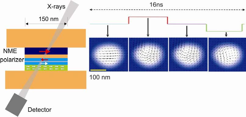
Development of Resonant Coherent X-ray Scattering
–
The scientific motivation for the development program
outlined below is the investigation of the critical
fluctuations of a magneticdomain structure at the
magnetic-paramagnetic phase transition. This research
falls into the general area of critical fluctuations at
phase transitions for which theory predicts that the
order parameter diverges at the transition temperature.
For magnetic phase transitions this implies that the
domain size should diverge,
i.e.,
right at the transition temperature a single magnetic
domain should extend –momentarily– over the entire
sample. However, this has never been observed
experimentally. One of the major reasons for this lack
of experimental proof is that impurities and defects
limit/influence these fluctuations. This limitation can
be overcome by studying critical fluctuations in
ultra-thin films with quasi 2-dimensional magnetization.
Such ferromagnetic films can be prepared essentially
defect free by epitaxial growth with thicknesses of only
a few monolayers. A further advantage of studying a thin
film is that the fluctuations are expected to be much
slower in thin films than in bulk materials. To
investigate the nature of these critical fluctuations,
the following four experiments will be undertaken:
-
Resonant Small Angle Scattering of Incoherent Soft
X-rays
Resonant scattering at the dichroic L3
absorption edge of magnetic transition metals will
yield statistical information about the magnetic
domain structure such as average domain size and
domain shape. Hence, when investigating the
temperature dependence of this statistical
information close to the transition temperature, the
average domain size of the critical magnetic
fluctuations can be derived.
-
X-ray Photon Correlation Spectroscopy (alias Dynamic
‘Light’ Scattering)
By
scattering of coherent photons, information about
the dynamics of the fluctuations can be derived from
the time dependence of the scattering intensities. A
third generation synchrotron light source like
SPEAR3 will enable time resolving dynamics occurring
on the microsecond time regime.
-
Ultra-Fast, High-Resolution, Lensless Imaging of
Magnetic Domain Structures
One potential application of SLAC’s upcoming X-ray
free electron laser LCLS will be ultrafast,
high-resolution lensless imaging. This will allow
recording of femtosecond snap shots of the magnetic
domain structure at and in the vicinity of the
magnetic phase transition. A series of such images
will enable distinguishing “real” magnetic
fluctuations from defect-pinned fluctuations.
-
Ultra-fast X-ray Photon Correlation Spectroscopy
At LCLS, a beam splitter and a delay line will be
used to obtain two femtosecond short X-ray pulses
separated by a variable delay ranging from a femto-
to a few picoseconds. Using both these pulses for
lensless imaging of the magnetic domain structure
will reveal the dynamics of the magnetization
fluctuations on a femto- to picosecond time scale.
Commissioning of the rebuilt BL5-2 continued, which also
included commissioning of the dedicated end station for
coherent soft X-ray scattering. Double pinhole test
scattering patterns were recorded to characterize the
coherence properties of the beam line. In addition, a
novel implementation for coherence measurements based on
a non-redundant array of pinhole structures was
developed and successfully applied.
The feasibility of phase contrast imaging in resonant
soft X-ray holography was demonstrated by imaging of a
magnetic domain structure. The important implication of
this achievement is that sample damage can be reduced
significantly by using phase instead of amplitude
scattering contrast.
In addition, multiple reference beam Fourier transform
holography has been developed. Since an image is
obtained simultaneously from each reference beam, the
effectiveness of the imaging technique increases
linearly with the number of reference holes. This allows
to further reduce the required dose for imaging on the
nano-scale, which is in particular important for
radiation sensitive samples like organic materials.
The thin film preparation chamber assembled during
FY2005 has been commissioned. First ultrathin magnetic
films for the investigation of critical magnetic
fluctuations have been grown. In preparation for these
experiments, temperature control, earth field
compensation, and an optical MOKE system to characterize
the magnetic properties of the thin films were
developed.
Small and Wide Angle Scattering Studies of Soft Matter &
Colloids
–
A proposal for a new SAXS/WAXS Materials Science beam
line at SSRL has been developed in response to the
burgeoning demand for the technique in a host of modern
nano-scale science applications. The beam line’s design
takes advantage of the accrued knowledge of the decades
of experimental scattering from synchrotron sources in
the field of materials science. Detailed plans
concerning its physical specifications and configuration
are already in place: with a double crystal
monochromator (interchangeable between silicon [111]
crystals and multilayers) and a horizontal and vertical
focusing toroidal mirror and five meter path length
downstream of the sample environment. This will provide
a beam ~1013
photons able to probe correlation lengths within
physical media up to half a micron in size at time
resolutions of 100 ms or less, thereby opening up a
facility for a whole new range of science. Some of this
new science that would be made accessible by this
geometry would include many pore size distributions
(spatially) and matrix complexations (temporally) that
are currently beyond the abilities of modern facilities.
Nanoparticles for Environmental Sorption Control.
This project focused on the nano-scalestructural
chemistry and environmental chemical dynamics of
bacteriogenic manganese oxide (MnO) nanoparticles.
Investigations at BL1-4 addressed the relationships
between particle size and MnO structure and stability
and the factors controlling the colloid chemistry of MnO
nanoparticles in aquatic systems. Insofar as Mn plays
unique and important roles in local and global elemental
cycles, including those of C, S, N, Fe and numerous
other trace metals, the knowledge gained from this
project will contribute towards our understanding of the
dynamics that control the chemistry of our soils,
natural waters, and atmosphere. Initial measurements of
bacteriogenic Mn oxide particle sizes in
bacteria/mineral mixtures were made allowing the
feasibility and test techniques for dispersing particles
to be assessed.
Reflection Geometry SAXS.
The capabilities of the new experimental hutch permitted
collection ofthe first SAXS data in a reflection
geometry, studying controlled growth of silicon
nanotubes intended for nano-scale thermoelectric
applications. In an effort to access the pores and
achieve significant alignment, a method was developed
for producing vertical pores out of the plane of a
substrate. Cubic mesoporous titania was believed to have
produced an ultra-flat hexagonal pattern from its 111
face that could be used to surface nucleate vertical
growth in the silica 2D hexagonal system SBA-15 and
hence the creation of accessible, aligned pores. Early
data, in the reflection geometry, permitted
characterization of the packing arrangement and aspect
ratio of these nanotubes, which revealed difficulties in
the synthesis, leading to misaligned tubular structures,
which remain to be corrected.
Nanoporous Metallic Media. Nanoporous metals were fabricated electrochemically by
selective dissolution or ‘dealloying’. When immersed in
a suitably aggressive reagent (e.g.
nitric acid), themore active metal was removed leaving
behind a ‘sponge-like’ bicontinous network of the noble
element. We have studied AgAu and CuPt alloys, which
formed nanoporous Au or Pt, respectively. SAXS has been
used to characterize the pore sizes and the pore
morphology as a function of dealloying time (1 minute to
3 days). We found that with increasing dealloying time
the average ligament spacing increases and the
morphology develops into a bicontinous network which
then coarsens. In a related study, diffraction has been
used to characterize the strain that develops during
dealloying.
Structural Properties of Novel Materials
–
Several major subgroups of materials are explored in
this area, including the local structure of
non-crystalline materials, thin films, and nanoporous
materials.
Non-crystalline Materials
–
A second experimental run was performed on the structure
of liquid water. Several experimental improvements were
made, including using an energy of 19.6 keV instead of
12 keV, which increases the maximum momentum transfer to
19.7 Å-1.
Secondly, asmaller, 600 micron orifice was used in the
water jet. The smaller water diameter results in better
energy resolution within the diffracted beam analyzer,
which allows for better resolution between the elastic
and Compton components of the diffracted beam. We also
implemented a thermal bath for the recirculated water
beam enabling us to measure the scattering both at room
temperature (23 C) and at 5 C, very close to the density
maximum of 4 C.
The analysis of the scattering from water is underway.
Some of the challenges needing to be met during the
analysis include the correct separation of the elastic
and Compton components of the scattered beam. The
advantage of increased energy resolution improves but
does not eliminate the overlap between the elastic and
Compton components. In a small region of momentum
transfer(roughly between 2 and 6 Å-1)
there is overlap of the two peaks. Analytical techniques
are being developed to consistently separate the two
components. A second challenge is the correct form of
the independent scattering. A molecular form factor has
been calculated for water vapor, but it is expected that
there will be subtle changes in the shape of the form
factor for liquid water. Weintend to use the excess
electron density at small atomic separation to modify
the form factor for liquid water. We plan to submit the
work for publication during this period as well as write
the doctoral dissertation.
Thin Film Structure
–
Studies on organic semiconductors have been extended to sexithiophene and substituted oligothiophenes, such as
p-tolyl-trithiophene-p-tolyl,
and studies will be initiated of other semiconducting
conjugated polymers, such as
poly[5,5’-bis(3-dodecyl-2-thienyl)-2,2’bithiophene],
PQT-12, which has a high field-effect mobility.
Nanoporous Metals
–
Nanoporous metals can be fabricated electrochemically by
selective dissolution, or ‘dealloying’. When immersed in
a suitably aggressive reagent, the more active metal is
removed leaving behind a ‘sponge-like’ bicontinous
network of the noble element. Alloys of interest include
AgAu, CuAu, CuPt, which form nanoporous Au or Pt for
example. X-ray
scattering methods (SAXS and GIXS) have been used to
carefully characterize the pores and the near-surface
structure in these materials.
Charge Density Wave Materials
–
Rare earth (R) tellurides (RTe2
and
RTe3)
are ideal materials in which to study charge density
waves (CDW), since the CDW gap is large and large
crystals can be grown. High-resolution X-ray diffraction
has been used to study the nature of the incommensurate
and commensurate CDWs in
RTe2
and
RTe3
with initially
R=La
and Ce. From the diffraction data, the atomic
displacements, which permit a deeper understanding of
the observed thermodynamic and transport properties of
these materials, have been obtained.
Ultra-trace and Microanalysis
–
A micro-focus X-ray mirror system using Kirkpatrick-Baez
(KB) optics was commissioned in the back hutch of BL6-2
and first real data studying interplanetary dust
particles were collected. Initially, this required a
profound understanding of the alignment of the newly
upgraded upstream beam line optics illuminating the
virtual source of the KB system. This adjustable virtual
source slit allows mapping to be carried out at variable
focal spot sizes between approximately 2 and 15 microns.
Photon fluxes fall in the 108
– 1010
photon/second range depending on the X-ray spot size, and
the accessible energy range covers readily the elements
Mg through Br. An aluminum-coated BN window allows
simultaneous imaging of the sample with a high
resolution optical microscope during the X-ray
measurements. A helium gas shower is mounted above the
sample to minimize radiation damage (due to ozone
generation) as well as spectrum “contamination” by argon
fluorescence from air. This shower creates a gentle flow
of He without introducing vibrations to the sample mount.
The X-ray hutch itself has been temperature-stabilized
minimizing possible X-ray beam and/or sample drift
during measurement. In addition, the data collection
software SUPER was upgraded allowing the collection of
the entire emitted fluorescence spectrum for each pixel
rather than collecting only predefined single channel
analyzer windows for selected elements of interest. This
is crucial in particular for studying samples containing
a variety of elements such as interplanetary dust
particles. In those cases overlapping fluorescence lines
in particular from neighboring elements can easily lead
to false images of the elemental distribution. Efforts
were made to evaluate different software packages for
quantitative analysis of the individual fluorescence
spectra. A software package developed at the ESRF called
PyMca is currently being employed for quantitative
analysis. The KB-based experiments in FY2005 were very
successful, given the short amount of beam time
available for characterization of the optical setup. In
addition to optics characterization, elemental maps and
trace element spectra were obtained for several thin
sections of micrometeorites. These samples included
round-robin samples provided by the Bulk Chemistry
subteam leader of the Stardust Preliminary Examination
Team. Our microprobe has been used in the preliminary
examination of the samples returned by the Stardust NASA
mission which swept through the tail of a comet (comet
Wild 2) resulting in the first successful sample return
mission since the Apollo program.
In FY2006, the KB optic-based microfocus facility has
opened up for external user groups. A first user group
from Canada has already been on line in January 2006,
studying Fe distribution in fruit fly brains as a model
for human Alzheimer disease. In addition, SSRL
scientists performed preliminary test measurements for
upcoming user groups studying Cu distribution in mice
liver as a model for the Wilson disease, as well as of
transition metal distribution in arthropod tools, such
as spider fangs or worm teeth.
In addition to the work on the microprobe, a new TXRF
chamber was designed, built and commissioned on BL6-2
mostly for studying small samples from the Genesis
mission. Despite its hard landing, this mission returned
fragmented samples of solar wind collected at the
Lagrangian point for over two years. TXRF, due to the
ability to distinguish between surface contamination
(from Utah desert) and the true implanted solar wind, is
an especially effective technique for studying these
types of samples. In particular, the sample holder of
this new setup was tailored for mounting small (cm2)
size samples without adding any contamination. In close
collaboration with the PI of the Genesis mission,
flight-spare samples and flown Sapphire samples have
been analyzed with high detection sensitivity. These
indicate that while some of the returned sample pieces
have significant surface roughness and contamination,
effectively reducing the sensitivity of the measurement,
others are sufficiently clean and smooth to enable us to
measure bulk solar
wind of dominant species (e.g.
Fe). Further examination of samples will determine
whether minority species will be able to be quantified.
Finally, the proposal to NIH (1 R01 EB004321-01) with
the title “A multi-keV X-ray microscopy facility for
bio-imaging” for development of a zone plate-based,
multi-keV Transmission X-ray Microscope (TXM) facility
was funded in May 2005 by the Bioengineering Research
Partnerships program of NIBIB. The total award was
distributed over four years and started in May 2005. The
purchase order for the basic TXM instrument from Xradia
Inc. was placed in September 2005. This instrument will
provide important and currently at SSRL unavailable
capabilities for the study of biological systems
in-situ,
such as the imaging of elemental distributions within
single cells or tissues with high spatial resolution.
The full field microscope will operate using photon
energies between 5 – 13 keV and exploits the advantages
of hard X-rays for 2D and 3D absorption and phase
contrast microscopy such as large penetration depth, a
large depth of focus, analytical sensitivity and
compatibility with wet specimens. Together this will
allow high resolution
in-situ
imaging, tomography and spectromicroscopy without
extensive sample preparation. The TXM will be delivered
from Xradia Inc. in October of 2006. We are currently
building a new X-ray hutch on BL6-2 for this
high-resolution microscope. It is anticipated that
commissioning will begin in fall of 2006 and by the end
of 2006 resolution test structures will be imaged in 2D
and 3D with a spatial resolution below 60 nm in
absorption contrast.
XAS Studies as a Probe of Electronic Structure /
Contribution to Function
–
X-ray absorption spectroscopy (XAS), at the ligand and
metal K- and metal L-edges, is used to determine the
electronic and geometric structure of metal-based
centers in inorganic and bioinorganic systems. The goal
is to understand the involvement of the metal center in
catalytic cycles important in industrial and biological
catalysis. The experimental approach is complemented by
Density Functional Theory (DFT) calculations,
photoemission spectroscopy measurements, valence bond
simulations and the development of data analysis tools,
as well as by other non-synchrotron-based methodologies
including MCD, resonance Raman and EPR spectroscopies,
where applicable. The combined approach enables
significant insight into geometric and electronic
structure and their contributions to reactivity.
Currently, studies are focused on systems containing
copper, iron, titanium, manganese, molybdenum, and
tungsten, with the anticipation of extending these
studies to other transition metal sites.
Copper –The
geometric and electronic structures of two mononuclear
CuO2
complexes [Cu(O2){HB(3-Ad-5-iPrpz)3}]
(1)
and [Cu(O2)(β-diketiminate)]
(2)
have been evaluated using CuK- and L-edge XAS studies in
combination with valence bond configuration interaction
(VBCI) simulations and spin unrestricted broken symmetry
density functional theory (DFT) calculations. These
systems are related to oxygen activation at a single
copper center, as in dopamine βmonooxygenase and
peptidylglycine α-hdroxylating monoxygenase. Cu K- and
L-edge XAS data indicated the Cu(II) and Cu(III) nature
of
1
and
2,
respectively. Total integrated intensity under the
L-edges showed that the ψ*LUMO
in
1
and
2
consists of 20% and 28% Cu character, respectively,
which is indicative of a very covalent ground state in
both complexes, although more so in
1.
This is consistent with VBCI simulations, which also
indicated that the ground state in
2
has more d8
n-character. DFT calculations showed that the ψ*LUMO
in both complexes is dominated by O2
character, although the O2n-
character is higher in
1.
It was also shown that the strong donor ligand in
2
is responsible for tuning the molecule towards more Cu(III)-peroxide like character in contrast to
trispyrazolyl borate in
1
which leads to a Cu(II)-superoxide species.
Heme-Copper Oxidases –
The geometric and electronic structure of the untethered
heme-peroxocopper model complex [(F8TPP)FeIII-(O22-)-CuII(TMPA)](ClO4)
(1)
has been investigated usingCu and Fe K-edge EXAFS
spectroscopy and DFT calculations in order to describe
its geometricand electronic structure. The Fe and Cu
K-edge EXAFS data indicated a Cu···Fe distance of ~3.72
Å. Spin unrestricted DFT calculations for the ST
= 2 spin state were performed on [(P)FeIII-(O22-)-CuII(TMPA)]+
as a model of
1.
The peroxo unit is bound end-on to the copper, and
side-on to the high spin iron, for an overall µ-η1:η2
coordination mode. The FeIII-peroxide
η2-bond
has two
components that arise from the donor interactions of the
peroxide π*σ
and π *v orbitals with the Fe
dxz
and dxy
orbitals, which give rise to σ and δ bonds,
respectively, while for the Cu site the
primary bonding interaction is between the peroxide
π*σ
and Cu dz2
orbitals. The π*σ
interaction
of O22-with
both Cu (η1)
and Fe (η2)
provides an effective superexchange pathway for strong
antiferromagnetic coupling between the metal centers.
Non-Heme and Heme Iron
Cyanides
–
Distinct spectral features at the Fe L-edge of the two
compounds K3[Fe(CN)6]
and K4[Fe(CN)6]
have been identified and characterized as arising from
contributions of the ligand π* orbitals due to
metal-to-ligand back-bonding Analysis of the L-edge
spectral shape, total intensity and energy shift have
been used to quantify the contributions of σ-donation
and π-back-donation to metal cyanide bonding. The
methodology developed demonstrates the application of Fe
L-edge XAS as a direct probe of metal-to-ligand
back-bonding.
Heme vs. Non-Heme Fe
–
Fe porphyrin compounds, or hemes, form the basis for
electron transfer in a number of biological systems,
with the most well-known being the cytochromes, which
effect electron transfer by shuffling between low spin
Fe(II) and Fe(III). The delocalization of the Fe
d-orbitals into the porphyrin ring has been difficult to
study spectroscopically because of the dominant
porphyrin π →π* transitions, which obscure the metal
based d-d bands. Recently, we have developed a novel
methodology that allows for the interpretation of the
multiplet structure of Fe L-edges in terms of
differential orbital covalency (i.e. differences in
delocalization of the different d orbitals) using a
valence bond configuration interaction (VBCI) model.
Applied to heme systems, this methodology allows
experimental study of the delocalization of the Fe
d-orbitals into the porphyrin ring. This methodology has
been applied to study two model systems
[Fe(tpp)(ImH)2]Cl
and [Fe(tpp)(ImH)2]
(low spin Fe(III) and Fe(II), respectively) and have
compared their multiplet structure to those of the two
low spin non-heme compounds
[Fe(tacn)2]Cl2
and [Fe(tacn)2]Cl3.
The Fe L-edge spectra are very sensitive to the effects
of π donation and π back-donation. The e1
hole (in heme,
D4h;
t2g,
in
Oh)
in the ground-state electronconfiguration of low spin
Fe(III), creates a dominant spectroscopic feature which
was quantified in
terms of
π
donation. The L-edge spectrum of [Fe(tpp)(ImH)2]
exhibits additional transitions caused by back-bonding,
which was also quantified. It was found that heme acts
as a substantial �-donor to Fe(III) but only a minimal
�-acceptor to Fe(II), indicating the electron transfer
involves a hole-based super-exchange mechanism.
Siderophores
–
In order to overcome the immense difference between
environmentally and nutritionally available Fe, many
microorganisms produce low-molecular weight
iron-chelators called siderophores. Our recently
developed L-edge methodology, described above, provides
a way of studying the bonding in these compounds. Fe
L-edge data on a small set of compounds,
K3[Fe(oxalate)3],
[Fe(pha)3]
and K3[Fe(catecholate)3]
have been obtained. Results show that both
the Fe K- and the L-edges shift to lower energy across
the series: K3[Fe(ox)3]<
[Fe(pha)3]<K3[Fe(cat)3].
This shift indicates a decrease in effective nuclear
charge and an increase in electron donation by the ligands. The total intensity of the Fe L-edge
transitions decreases across the series,
implying that the Fe-O bonds of K3[Fe(cat)3]
are the most covalent of the compounds. By simulating
the shape of the spectra, it has been possible to
calculate the σ and π contributions to bonding in the
compounds, providing insight into the factors which
affect the differences in their thermodynamic stability.
Iron-Sulfur
–
Ligand K-edge XAS has been used to obtain a quantitative
description of Fe-S bonding in Fe-S model complexes and
protein active sites. The results of these studies have
been correlated to the extent of H-bonding and
differences in redox potentials. To develop a
quantitative description of hydrogen bonding in Fe-S
systems, a series of P450 model complexes, where the
amount of hydrogen bonding was systematically varied,
were examined by S K-edge XAS. The data show a dramatic
decrease in pre-edge intensity with increasing H-bonding
to the ligated thiolate. DFT calculations reproduced
these effects and showed that the observed changes are in
fact solely due to H-bonding to the thiolate ligand. The
energy of the H-bonding interaction was estimated to be
-2.5 kcal/mol in the gas phase. The rather small
H-bonding energy appears to contrast the large change in
ligand-metal bond covalency (30%) observed in the data.
A bond
decomposition analysis of the total energy was developed
to correlate the pre-edge intensity change to the change
in Fe-S bonding interaction on H-bonding. This analysis
showed that the Fe-S interaction energy is greatly
reduced due to H-bonding. This effect is greater for the
reduced than the oxidized state, leading to an ~350 mV
increase in the redox potential. It was found from a VBCI
model that Eo
should vary linearly with the covalency of the Fe-S bond
in the oxidized state, which can be determined directly
from S K-edge XAS. The above study was extended to a
hydrogen bonded [Fe4S4]2+
cube, which had an elongated core structure in contrast
to the compressed
core structures of most [Fe4S4]2+
cubes. A decrease in pre-edge intensity was observed for
the H-bonded cube. DFT calculations indicated that the
change in Fe-S covalency observed experimentally had
almost equal contributions from the cluster elongation
and the H-bonding
interaction. These calculations also indicated that the
elongation of the [Fe4S4]2+
cube changes the spin topology of the ground
state due to redistribution of the ligand superexchange
interactions in the cluster.
The geometric and electronic structure of the active
site of the nonheme iron enzyme nitrile hydratase
(NHase) has been studied using S K-edge XAS and DFT
calculations. Using thiolate (RS-),
sulfenate (RSO-)
and sulfinate (RSO2-)
ligated model complexes to provide benchmark spectral
parameters, the results showed that the S K-edge XAS is
sensitive to the oxidation state of S-containing ligands
and that the spectrum of the RSO-species
changes upon protonation as the SO bond is elongated (by
~0.1 Å). These signature features were used to identify
the three cysteine residues coordinated to the low-spin
FeIII
in the active site of NHase as CysS-,
CysSOH andCysSO2- in
both the NO-bound inactive form and the in the
photolyzed active form. These results were correlated to
geometry optimized DFT calculations. The pre-edge region
of the XAS
spectrum is sensitive to the Zeff
of the Fe and revealed that the Fe in the [FeNO]6
NHase specieshas an effective nuclear charge very close
to that of its photolyzed FeIII
counterpart. DFT calculations revealed that this results
from the strong π back-bonding in to the π anti-bondingorbital
of NO, which shifts charge from the formally t26
low-spin ferrous center.
Titanium Cyclopentadienyl Catalysts
– Ti-TEMPO complexes (TEMPO =
2,2,6,6tetramethylypiperidine-N-oxyl) provide a means
for generating Ti(III) complexes by homolysis of the
Ti-O bond. The rate of Ti-O bond homolysis depends on
the ancillary ligation to the titanium, and it has been
determined that bis-Cp-Ti-TEMPO (Cp=cyclopentadienyl)
complexes readily undergo homolytic cleavage, while the
mono-Cp-Ti-TEMPO complexes do not. Recently, Ti K- and
Cl K-edge XAS studies have been applied to a series of
Ti-TEMPO complexes
(TiCl3TEMPO,
TiCl2CpTEMPO,
TiClCp2TEMPO).
The XAS results indicate that these complexes are best
described as Ti(IV)-TEMPO anions. The Cl K-edges show
that the replacement of Cl by Cp weakens the remaining
ligands, demonstrating a spectator ligand effect which
is one factor that contributes to Ti-TEMPO bond
homolysis. However, correlation of the XAS results to
DFT calculations shows that stabilization of the
three-coordinate product by Cp makes a more significant
contribution to the energetics of Ti-O(TEMPO) bond
homolysis.
PES Studies of Electronic Structure Contribution to
Function
– The shake-up satellite structure present in core and
valence photoemission spectroscopy (PES) data is being
used in combination with a valence bond configuration
interaction (VBCI) model to experimentally quantify
electronic relaxation (i.e. the change in electronic
structure of metal complexes upon oxidation) and its
contributions to reduction potentials and kinetics of
electron transfer. Variable-energy PES (VEPES)
experiments provide the tool to maximize the metal
contribution, while minimizing that of the ligand, to
the valence band region through cross section effects
(delayed
maximum and Cooper minimum) and resonance enhancement.
VEPES data on a series of model iron complexes – high
spin [FeCl6]4-/3-
and low spin [Fe(CN)6]4-/3-,
[Fe(tacn)2]2+/3+,
[FeTpp(HIm)2]1+/0,
[FeTpp(Py)2]1+/0
– were measured and analysis initiated.
Materials Research
Research carried out by SSRL faculty and staff and
associated Stanford faculty and students covers a broad
set of disciplines: (1) Complex Materials; (2) Magnetic
Materials; (3) Scientific and Educational Gateway
Program (through FY2006); (4) Novel Materials and Model
Systems for the
Study of Correlated Phenomena; (5) Nano-scaled Magnetism
in the Vortex State of High-Tc
Cuprates; (6) Nano-scale Electronic Self-Organization in
Complex Oxides; (7) Nano-Magnetism;
(8) Behavior of Charges, Excitons and Plasmons at
Organic/Inorganic Interfaces; and (9) Development and
Mechanistic Characterization of Alloy Fuel Cell
Catalysts. Areas (4) through (8) are collaborative
efforts of the SSRL X-ray Laboratory for Advanced
Materials and the Stanford University Geballe Laboratory
for Advanced Materials.
1. Complex Materials
Z.-X. Shen, S. Doniach, M. Greven, R.B. Laughlin, X.J.
Zhou, K. Tanaka, H. Li, L. Lu, K.Downum, Y. Cho, J.
Hancock, D. Santiago, D. Schroeter, L. Zhou, X. Zhou,
B.A. Bernevig
The team has conducted comprehensive experiments using
photoemission (Shen), scattering (Greven and Shen), and
theoretical investigation (Laughlin and Doniach) on
complex materials, and has made substantial progress in
this period. There also are synergetic interactions with
the nano-science programs of Greven, especially in the
area of single crystal growth, and Shen’s core program
on strongly correlated materials.
A major focus of the program is the electron-doped
materials that have presented an important challenge to
the systematic understanding of high-temperature
superconductors. The Greven group
has continued its successful effort on the
electron-doped superconductor Nd2-xCexCuO4
(NCCO), built on its early success in growing
high-quality single crystals. Recent results include the
first determination of spin-correlations in
superconducting samples, which constitutes significant
progress toward a full understanding of the normal state
of the electron-doped superconductors.
Since superconductivity in high-Tc
cuprates appears in close proximity to the
antiferromagnetic phase, it is essential to understand
the nature of nearby magnetic ground states. Through
careful X-ray and neutron diffraction work, Greven
discovered that the oxygen reduction process, required
to render NCCO superconducting, transforms a fraction of
the crystals into cubic
(Nd,Ce)2O3,
and that the field-effects observed by others and
ascribed to a quantum phase transition of NCCO are not
intrinsic, but due to this secondary phase.
Consequently, the question of genuine magnetic field
effects in NCCO has remained a very interesting,
unresolved research topic. By studying the magnetic
field effect on the spin excitations, Greven was
recently able to obtain new data consistent with the
absence of field-induced magnetic order [Motoyama
et al.,
preprint]. Moreover, these measurements constitute the
first neutron scattering study of the effects of a
magnetic field on the superconducting magnetic gap in
NCCO. The discovery of spurious magnetism in NCCO is a
good example of the benefits of a synergistic growth and
scattering effort like that by Greven.
Greven continued the novel use of inelastic X-ray
scattering to investigate the collective charge
excitations in the model high-temperature superconductor
Hg1201, the single-layer material with the highest value
of Tc,
and of the parent compound La2CuO4.
This latter work, carried out at the APS and made
possible by newly available large Hg1201 crystals grown
by Greven, led to the discovery of a remarkably rich
structure of electron-hole pair excitations in the
cuprate superconductors.
Greven's work on the structural phase diagram and
charge-order phenomena in the layered manganite was
extended to cover a wider range of doping as well as
neutron scattering. These results will allow a
comprehensive understanding of the structural and
magnetic phase diagram.
The angle-resolved photoemission spectroscopy (ARPES)
component of the program (Shen) has two primary tasks,
research, and operation of the beam line 10.0.1
end-station at the ALS in support of this research. That
activity also benefited a broader community performing
ARPES experiments using the end-station.
The focus during this period of time is to understand
the nature of collective modes coupled to cuprate
superconductors. Data with very high signal-to-noise
level, together with a numerical method developed in
collaboration with Ward Plummer’s group (Oak Ridge
National Laboratory), enabled a glimpse of the phonons
and their coupling to electrons in cuprates. This
represents the first experimental evidence for
multi-boson coupling in cuprates.
Another area of major progress during the period is the
new insight on the polaronic behavior of single hole in
undoped cuprate. Based on ARPES data from La2CuO4
and phonon spectra fromneutron scattering, theoretical
calculation by Gunnarsson’s group (Max Plank Institute)
show that the spectra are consistent with the material
being in the polaronic regime, which is consistent with
our earlier finding for copper oxide chloride.
Shen’s group also has made important technical progress
in applying synchrotron radiation for high resolution
spectroscopy experiments. The resolution and flux
density at the ALS have been a critical factor for the
program in generating a significant database. On the
other hand, this also raises the technical problem of
space charging. Shen’s group has systematically
characterized this problem and our finding is generally
useful in guiding high resolution photoemission
experiments in third generation synchrotron radiation
facilities.
A major advance by Shen’s group during this period is
the discovery of nodal quasiparticle and nodal antinodal
anisotropy in layered colossal magnetoresistive
manganites. This finding providesdeep new insights on
the “pseudogap” phenomena in cuprates. The pseudogap
behavior has for a long time been considered to exist
only in superconducting materials, and thus is directly
related to high-temperature superconductivity. Finding
this same behavior in ferromagnetic state suggests that
the relationship between “pseudogap” and
superconductivity is more subtle. In particular, this
finding supports the notion that there are two
“pseudogaps”, and the larger pseudogap is in fact a
reflection of a competing state to superconductivity.
Thus, both superconducting and pseudogap phases are
manifestation of the same underlying physics that give
rise to the rich and intricate phenomena seen in these
complex oxides.
The work of the Laughlin group in 2005 was conducted
mainly by David I. Santiago and Zaira I. Nazario. The
Laughlin group has studied the phase diagram in cuprates
high Tc
superconductors.Recent experimental measurements suggest
the coexistence of various phases of matter in different
regions of the pseudogap regime. Guided by such
experiments the group has studied the properties of a
spin-density-wave antiferromagnetic mean-field ground
state with d-wave superconducting (DSC) correlations.
This work concentrates in the case when
antiferromagnetic order is turned on weakly on top of
the superconductivity, which corresponds to the onset of
antiferromagnetism at a critical doping. In such a case
a small gap proportional to the weak antiferromagnetic
gap opens up for nodal quasiparticles, and the
quasiparticle peak would be discernible.
The program looked broadly at the many-body problem in
condensed matter system, beyond the
high-Tc
superconductors and transition metal oxides. Work on the
superfluid to Mott insulator transition in bosonic
systems has been done, where the phase diagram of a
single component Bose Einstein Condensate (BEC) in an
optical lattice at zero temperature was obtained. In
that work, the discontinuous nature of the transition
between the superfluid and the Mott insulator (under
certain conditions) was elucidated, as well as its
independence on commensuration of the number of bosons
with the lattice. Recently, measurements which could be
interpreted as such a transition have been performed by
Mark Kasevich's group of Stanford University. While,
being superfluids, BECs share many properties with
superfluid helium, they have never been seen to share
the existence of roton excitations which are present in
helium. Superfluid helium is close to becoming a solid.
The roton minimum is a consequence of enhanced density
fluctuations at the reciprocal lattice vector of
the stillborn solid. Thus rotons have not been observed
in BECs in atomic traps since they are not near a solid
phase, but if they are tuned near a transition to a Mott
insulating phase, a roton minimum will develop at a
reciprocal lattice vector of the lattice. Equivalently,
a peak in the structure factor will appear at such a
wave vector. The smallness of the roton gap or the
largeness of the structure factor peak are experimental
signatures of the proximity to the Mott transition.
Laughlin’s group has also done some work on
metal-insulator transitions. Specifically, the work was
motivated by V2O3
and f-electron systems which have phase diagrams in
which a line of first order metal-insulator transition
ends at a critical point above which the two phases are
indistinguishable. Bob Laughlin's Gossamer technique was
extended to show that the Gossamer metal in a single
band model will describe a metallic phase that becomes
arbitrarily hard to differentiate from an insulator as
one turns the Coulomb correlations up. Thus one can go
continuously from the metal to the insulator.
Laughlin’s group also has done some work on quantum
criticality that should be published this year. In
collaboration with Jan Zaanen, they have an article on
spin-orbit coupling and interesting quantum interference
effects that will also be published in this period. The
Doniach group expects to continue work on the relation
between superconductivity and electron correlations in
intercalated graphite. Recent work on superconductivity
in doped graphite intercalated compounds has suggested
that, despite theoretical work to model this by
conventional electron phonon coupling BCS theory, these
theories have been unable to explain why different
intercalates have
very different Tc’s.
Tc
spans at least one order of magnitude when going through
different intercalates and it is believed possible to
study the relative effects of t-J correlation coupling
versus electron-phonon coupling on these various Tc’s.
The Doniach group has been focusing on looking at
electron correlations in graphite. Although this has
traditionally been considered in the context of band
theory, recent experiments by Kopelevich and
collaborators show strong evidence of a semi-metal -
insulator transition in applied magnetic fields of order
1 kG. The experimentalists found that a scaling relation
suggested by Das and Doniach in 2001 for the
superconductor – “bose metal” state of thin
filmsuperconductors also applies to the graphite data.
The effects of correlations in graphite have been
studied using a t-J model, following ideas proposed by
Baskaran for the superconductor MgB2.
2. Magnetic Materials Research
J. Stöhr, H. Siegmann, H. Ohldag, Y. Acremann
The general goal of this program is to develop new
techniques and approaches for the study of modern
magnetic materials in the form of thin films,
multilayers and nanostructures and explore the origin of
magnetic phenomena associated with such materials.
Work has continued on the phenomenon of
exchange bias
by means of X-ray microscopy and spectroscopy. In
particular, soft X-ray dichroism absorption spectroscopy
was used to investigate
the direction of interfacial exchange coupling in a
ferromagnetic / antiferromagnetic Co/FeF2 bilayer. This system behaves quite differently from
conventional exchange bias and, depending on sample
preparation, it can exhibit either positive or negative
exchange bias. Two different kinds of
interfacial uncompensated Fe moments were found in FeF2.
A smaller pinned portion couples antiparallel to the
ferromagnet and can lead to positive or negative
exchange bias depending on the size of the cooling
field. A larger portion couples more strongly and
parallel to the ferromagnet. It
increases the degree of antiferromagnetic order in FeF2
near the interface and plays an important role in the
observed coercivity increase at high temperatures. The
work was recently published: H. Ohldag, H. Shi, E.
Arenholz, D. Lederman and J. Stöhr,
Phys. Rev. Lett.
96,
027203 (2006).
The program on ultrafast magnetic switching with
electron beams and the program on lensless magnetic
imaging with X-rays, reported last years, have now been
funded separately as part of the Stanford Ultrafast
Science Center (PULSE) and the work is reported under
that program.
A new subprogram has started, based on the exploration
of new ways of exciting magnetic materials by ultrafast
magnetic field pulses. Rather than using electron beams
ultrafast laser pulses have been explored for launching
fast and strong current pulses. The idea is to generate
ultrafast magnetic field pulses by current flow through
a strip-line, as illustrated in Figure 1. The current
pulse is triggered by an ultrafast laser which either
opens an electro-optical (Auston) switch or creates
ultrafast photoemission into vacuum on one side of the
stripline which is grounded on the other side. Such a
scheme has the advantage of being compatible with
ultrafast X-ray imaging.
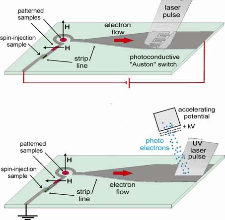
Figure 1. Arrangement for generating short and strong
current pulses traveling down a strip line. In the upper
picture the current pulse is generated by a
photoconductive switch, in the lower picture by a
photoemission pulse.
The scheme illustrated in Figure 1 using laser pulse
probe experiments will be explored and will characterize
the achievable pulse length and field strength. Samples
will be fabricated with our new magnetic thin film
deposition system and by use of lithographic
manufacturing techniques.
The design of a scanning transmission X-ray microscope
for BL13 at SSRL will also continue.
3. Scientific and Educational Gateway Program
A. Nilsson, A. Mehta and R. R. Chianelli
This continuing, joint effort with the University of
Texas at El Paso (UTEP) serves both the Mexican-American
and Mexican communities in undergraduate and graduate
education by engaging student scholars in science and
engineering research programs at all levels. The program
provides travel support for Mexican-American and Mexican
students and supporting faculty, science and
technological support by an SSRL scientific staff
member, who also assists participants in beam line
operations and laboratory facilities use, and a
scientific staff member at UTEP, who develops and
implements computational tools and software for analysis
of synchrotron data. These staff members train students
in methods of data reduction and analysis and, jointly
with SSRL staff scientists, develop collaboratory tools
for remote access to instrumentation and data measured
at SSRL. This program has been quite effective, as shown
by the number of UTEP students participating in
FY2001-2005 (almost 50 students). These students and
staff underwent training and carried out experiments on
existing SSRL peer-reviewed proposals coordinated across
five separate beam lines. The students continue to
enhance their training by taking highly successful
proposal writing classes at UTEP (Chianelli). SSRL staff
work closely with the UTEP faculty and staff to train
and support the new students and their research efforts.
Part of the period in FY2005 saw the synchrotron
start-up for the now successfully completed SPEAR3
upgrade. In addition, an electrical accident further
delayed the schedule. However, this allowed time for the
students to concentrate on data analysis with the help
of SSRL and UTEP staff. Progress during this period was
excellent with many students and faculty further
developing their proficiency in the use of synchrotron
techniques and producing publications based on previous
collected data. Several students achieved in-depth
understanding of synchrotron radiation and its
application by attending the Stanford-Berkeley Summer
School.
The level of synchrotron related skills increased
substantially for both the students and faculty, which
will be reflected by refereed publications and meeting
presentations of the group. Particularly skills were
improved in the areas of SAXS and carbon edge XANES.
Progress was made in six core areas in FY2005; (1) The
Chianelli group continued to address structure/function
relations in transition metal sulfide (TMS)
hydrodesulfurization (HDS) nanocatalysts. The first
“Gateway” Ph.D. (Myriam Perez) graduated and accepted a
post-doctoral position at SSRL; (2) Diffraction studies
on Maya Blue demonstrated that the pigment is a surface
complex of the indigodye with palygorskite clay; (3)
Data analysis on both the SAXS and WAXS data from
asphaltenes, crude oils from Mexico and Venezuela and
TMS catalysts was completed; (4) The Gardea group
continued to investigate the metal binding properties of
the “hyperaccumulators”; (5) The Pingitore group
continued to study trace elements in human bone. The
incorporation of Sr, Zn, Pb,
etc.
in human bone is a topic that impacts archaeology,
nuclear waste/terrorism, biomedicine, and environmental
pollution. All of these projects yielded significant
publications in 2005 and in press for 2006.
The SPEAR3 installation allowed time to develop the full
scientific impact of the program. This is shown by the
significant increase in the demonstrated impact of
synchrotron studies on the scientific understanding of
the problems discussed above. Students and faculty are
well trained to participate in “bringing on line” the
new SPEAR3 experiments and testing the new significant
capabilities.
Approximately 60 students will have been involved in
synchrotron programs at SSRL involving multiple
techniques, diffraction (SAXS and WAXS), scattering
techniques including anomalous dispersion scattering,
XAFS (hard and soft X-ray) and protein crystallography.
Many of these students will become a regular part of the
synchrotron user community and some will have
synchrotron use become a major part of their careers.
Many these students will have obtained experience at
other DOE facilities. Up to four students will have
completed or nearly finished a Gateway Ph.D. and become
full members of the synchrotron community. Several
faculty members at UTEP and related Mexican institutions
will have become new synchrotron users. Major scientific
progress will have been reported from the Gateway
program as discussed above. The progress will be of such
quality that the program will not only have made a major
impact on training minority scientists but the students
will have produced significant and competitive
scientific contributions.
4. Novel Materials and Model Systems for the Study of
Correlated Phenomena
I.R. Fisher, M.R. Beasley, T.H. Geballe, M. Greven,
N. Barisic, G. Chabot-Couture, A.S. Erickson, G. Koster,
L. Litvak, Y. Matsushita, N. Ru, P. SanGiorgio, K.Y.
Shin, G. Yu, X. Zhao, X. Zhou
This program directly addresses scientific questions at
the heart of understanding correlated electron behavior
in complex materials. This work explores the conditions
of occurrence and mechanisms behind these effects, and
examines the consequences for bulk properties and
collective phenomena. The approach is to identify and
synthesize model systems that enable exploration of
particular interactions in isolation. The essence of the
proposed research therefore lies in the design, growth
and characterization of novel materials.
The original title of this subtask was “Nanoscale
ordering in complex oxides: model systems for local
probes.” The title was updated during the recent renewal
to better reflect the focus of our current research.
Negative-U Impurities --Over
the course of the last three years a multistep synthesis
route has
been established to produce high-quality single crystals
of the unusual superconductor (Pb1-xTlx)Te,
and work has gone forward on detailed measurements to
characterize the thermodynamic, transport and
spectroscopic properties of these samples. The Tl
impurities appear to act as negative-U centers in this
material, and this research explores the role that these
impurities play in both the superconducting and normal
state properties. Early measurements established that a
critical concentration of Tl impurities is required to
cause superconductivity in PbTe. For Tl concentrations
beyond this critical value the Fermi level appears to be
pinned, such that the Tl impurities act in a
self-compensating manner. It seems that the
superconductivity is intimately linked to the presence
of a mixed Tl valence, the presence of which also is
reflected in the normal state transport of the material.
In particular, during FY2005 evidence was found for a
charge Kondo effect associated with degenerate valence
states of the Tl impurities. This observation, supports
the notion of an electronic pairing mechanism in
superconducting Tl-doped PbTe,
perhaps accounting for the unusually high Tc
value. Ongoing research seeks to further investigate this
effect in Tl-doped PbTe and related materials, testing
various predictions of the theoretical models that have
been proposed.
In FY006, the superconducting properties of Tl-doped
PbTe have been explored in more detail. Heat capacity
measurements for the highest Tl concentrations indicate
that the superconducting
anomaly is close to the BCS result (ΔC/γTc
~ 1.43), which within the charge Kondo model would imply
that the renormalized electrons are indeed participating
in the superconductivity. However, this quantity appears
to show a marked decrease as the impurity concentration
decreases, the origin of which effect is uncertain and
warrants further investigation. Additional insight can
be expected from a close collaboration with C. Gough and
E. M. Forgan (Birmingham, UK) exploring the penetration
depth via microwave conductivity and muon spin rotation.
Initial experiments from early this year indicate that
the T-dependence of the superfluid density is close to
BCS predictions, but with some unusual field dependence.
The continuing normal state magnetotransport
measurements show an unexpected trend towards a linear
T-dependence of the resistivity that we are continuing
to study. These data are currently being modeled by J.
Schmalian (Ames Laboratory) and P. Coleman (Rutgers) in
terms of a pair-diffusion process associated with the
negative-U properties of the Tl-impurities. Initial
EXAFS measurements (in collaboration with
F. Bridges, UCSC) are suggestive of two Tl-Te distances,
consistent with the proposed disproportionation, but are
limited in resolution due to the proximity of a nearby
Pb absorption edge. Experiments are also planned at the
NHMFL Microkelvin Laboratory (with Yoon Seuk Lee,
U. Florida) to look for reentrant behavior at low
temperatures, a key prediction of Schmalian’s charge
Kondo model. Additional external collaborations will
probe the effect of pressure on the superconducting and
normal state properties (S. Brown, UCLA), the optical
conductivity (Z. Schlesinger, UCSC) and core level
spectroscopy (C. Fadley, UCSD).
Charge-Density Waves in Layered Rare Earth Tellurides
-- We recently identified the
layered rare earth (R) tellurides RTe3
and RTe2
as model systems to study Fermi surface reconstruction
in incommensurate CDW compounds, a topic that has
significant bearing on current questions in the field of
cuprates and other strongly correlated oxides. These are
particularly attractive model systems because (a) the
electronic structure is especially simple and the band
filling can be tuned by chemical substitution; (b) the
CDW gap is large and can be measured by a variety of
powerful techniques; (c) we can explore the consequences
of the CDW formation on the magnetic properties of the
material; and (d) we are able to grow high-quality
single crystals, enabling sensitive probes of the
Fermi surface including dHvA and ARPES, and real space
probes including STM.
Initial angle resolved photoemission experiments in
collaboration with Z.X. Shen clearly revealed the CDW
gap, showing gapped and ungapped regions of the Fermi
surface. Our ongoing
experiments further explored the Fermi surface nesting
and CDW formation in both RTe3.
Further
experiments probing the FS reconstruction in RTe3
were conducted by a student supported by thisgrant
during an extended visit to A.P. Mackenzie’s research
group in St. Andrews, Scotland. These measurements
revealed multiple frequencies all of which vary with
angle according to 1/cos(theta), consistent with minimal
z-axis dispersion. The measurements are currently being
compared with ARPES data with the aim of fully
determining the reconstructed FS. Other experiments have
been performed at SSRL, following award of beam time.
Initial high-resolution diffraction experiments have
revealed an extremely well-ordered modulated structure
with minimal harmonic content. These measurements are
continuing with the ultimate goal of finding a model for
the atomic displacements in the CDW state, and hence
determining whether the material consists of short
regions of commensurate modulations with regular
discommensurations, or is indeed a truly incommensurate
structure.
Experiments at SSRL have established that the CDW in RTe3
has remarkably little harmonic content. Work is
currently in process to model the atomic displacements
in an effort to differentiate between a truly
incommensurate distortion and a modulation consisting of
a dense array of discommensurations. Further diffraction
experiments were planned to follow the T-dependence of
the CDW wave-vector, motivated in part by theoretical
models of S. Kivelson (Stanford) predicting a possible
phase transition between unidirectional (stripe) and
bidirectional (checkerboard) order. Additional
experiments and analysis will finalize our dHvA study,
while magnetotransport measurements will pursue our
recent observation of superzone gap formation in
the heavy rare earth members of the series (i.e.
an additional gapping of the FS at TN).
Work has
also continued to explore the role of Te vacancies in
RTe2-x
in determining the optimal nesting wave-vectors and
resulting CDW modulation in this single-layer variant.
Most excitingly, there is
now confidence the alternating single and double layer
compound R2Te5,
intermediate between the
single layer RTe2
and bilayer RTe3
compounds has been successfully synthesized. Experiments
are probing how band filling affects the CDW formation
in this family of compounds, providing a useful window
on the role of dimensionality (stripe vs checkerboard)
in CDW systems. Numerous internal and external
collaborations continue to probe other physical
properties of these materials, including STM (A.
Kapitulnik, Stanford), optical conductivity (L.
DeGiorgi, ETH, Zürich), NMR
(V. Brouet, Université de Paris Sud, France) and
positron annihilation (S. Dugdale, Bristol, UK).
Ferromagnetism in the Mott Insulator Ba2NaOsO6
-- The interplay between orbital and spin degrees of
freedom can play a pivotal role in many correlated
electron effects. Perhaps the clearest example of this
is to be found in ferromagnetic Mott insulators – a
rarified and unusual class of materials. In a single
band Hubbard model, hopping between adjacent sites leads
to anantiferromagnetic ground state. However, orbital
degeneracy can favor ferromagnetic exchange due to the
additional energy that can be gained from Hund's rule
coupling of electrons in two different orbitals. In such
a case, the appearance of ferromagnetism is thought to
be accompanied by a complex form of anti-ferro orbital
order, the precise nature of which depends strongly on
details of the specific lattice and material. This is an
exciting and topical area in correlated electron
physics. However, there are precious few candidate
materials. Furthermore, those that have been studied
belong exclusively to the 3d transition series.
Ba2NaOsO6
presents a wonderful opportunity to study the interplay
of orbital and spin ordering in the unusual environment
of a 5d1
ferromagnet. A method has recently been determined to
grow relatively large single crystals of this material
that enable a host of exciting experiments. Initial
thermodynamic and transport measurements, performed
early in FY2006, demonstrate that this material is
indeed a ferromagnetic Mott insulator, but with an
ordered moment that is substantially less than 1 μB
(i.e.,
the ordered state consists of a complicated spin
structure with a net ferromagnetic moment). The reason
for this is becoming evident. At room temperature,
Ba2NaOsO6
occupies an undistorted double-perovskite structure, in
which the OsO6
coordination polyhedra are neither rotated with respect
to the lattice, nor distorted from a perfect octahedral
symmetry. In this case, the three t2g orbitals are indeed degenerate, and ferromagnetic
exchange can be anticipated. However, the FCC lattice
cannot be decorated with three "color" orbitals in such
a way that each site always has nearest neighbors of a
different color. For adjacent sites of the same color
the superexchange is perforce antiferromagnetic. Hence,
in the absence of any additional structural phase
transition, it can be anticipated that the resulting
ground state of
Ba2NaOsO6
is a complex mixture of spin and orbital order, with a
strong ferromagnetic
component, but which falls short of 1
μB.
An active proposal is in place with the Advanced
PhotonSource (together with Z. Islam, APS, ANL) to study
the spin and orbital ordering in this compound later in
the year. Other ongoing experiments will probe the
high-field magnetization (with L. Balicas, NHMFL),
exploring the possibility of a metamagnetic transition
to the fully saturated state.
Electronic Inhomogeneities in Correlated Electron
Materials --
One of the goals of this programis to synthesize model
systems for the study of nanoscale electronic
inhomogeneities that result from the coulomb
interactions in correlated electronic materials, and
ultimately to study these inhomogeneities using scanning
probes. Toward this end, thin films of the Mott
insulator CuO have been grown, in which such nanoscale
inhomogeneities have been previously reported. This is
perhaps the simplest conceivable copper oxide related to
the high temperature cuprate superconductors. Epitaxial
thin films of stable monoclinic CuO have been grown
previously on MgO substrates. More recently CuO has been
grown epitaxially with square symmetry in the plane (i.e.,
either cubic or tetragonal overall). The composition is
confirmed by UPS spectra taken
in-situ
in an adjacent chamber. The structure has been confirmed
using
in-situ
RHEED and byPhoto-Electron Diffraction. Ion-beam
assisted deposition (IBAD) has been tried to further
enhance our ability to get a cubic structure, but this
has not so far been successful, due presumably to
differential sputtering of oxygen and cupper. Very
recently, it has also shown that CuO can be grown using
Pulsed Laser Deposition. The detailed properties of
films grown by these two methods now need to be
compared.
In parallel with this effort, thin films of ZrO2
have been deposited using pulsed laser deposition(PLD)
for use as high-K dielectrics for field-effect doping
applications. The student working on this project has
left the university. In light of this, focus will revert
to the CuO work and this part of the project will not be
continuing.
A square-planar (rock salt) form of CuO has now been
deposited successfully and physical study of these films
is being pursued. Standard transport and optical
measurements are being carried out, and various scanning
probes are being applied in collaboration with
colleagues here at Stanford. These include Scanning Hall
probes, MFM, STM and scanning potentiometry to look for
inhomogeneities. In addition, on the materials side,
studies will begin on approaches to dope these films.
This might be done through introducing oxygen deficiency
or by using strongly electropositive (or negative)
over-layers.
Superconductivity and Magnetism: Pair Density Waves and
SrRuO3 --Studies
of the superconductor/ferromagnetic proximity have drawn
a remarkable level of interest, primarily because it is
proving to be an effective model system for the study of
the interaction between superconductivity and magnetism.
One of the most striking predictions from theory is the
existence of decaying pair density waves in the
ferromagnetic side of an SF proximity bilayer due to the
exchange field in the F material. Evidence for this
effect is mixed. In order to get more detailed
information regarding SF proximity structures, in
collaboration with the group of Kookrin Char at Seoul
National University, tunneling density of states studies
are in progress on the F side of Nb/permalloy SF
bilayers as a function of the thickness of the F layer.
It is found that the striking result that the tunneling
density of states as a function of thickness (viewed as
deviations from the normal density of states) is of
constant form with amplitude that scales exponentially
over four orders of magnitude as a function of the F
layer thickness. No evidence for pair density
oscillations was found. More recently, a detailed
comparison of these results with the available
theory has been carried out. It is found that the theory
can account for these data only if patently unreasonable
values are used of the exchange field and the spin-flip
scattering rate. This work has now been accepted for
publication. One possible reason for this serious
discrepancy is that we are using very strong
ferromagnets whereas the theory assumes weak magnetism.
In order to clarify the situation, a second strong
ferromagnet (Ni) is presently being studied for the F
material.
Thin films of SrRuO3,
a rare example of a 4d itinerant ferromagnet continue o
be deposited and studied. With the group of Lior Klein,
the magneto-transport in SrRuO3
is being examined to test recent theories of the effect
of the Berry phase in magnetic systems. We did not
confirm the predictions. Studies also continue of
various physical properties of the films including spin
accumulation at domain walls during electric transport,
the magnetic anisotropy of the material and the
superconducting properties of Nb/SrRuO3
SF hybrids. In SF hybrids there is only magnetic coupling
between the S and F layers (i.e. there is no proximity
effect coupling).
Given
present results on the tunneling density of states on
Nb/permalloy SF proximity bilayers, it is very important
to study other F materials to see if these results are
universal or specific to permalloy. Studies with Ni as
the F material were mentioned above. The next obvious
step would be to move systematically to lower exchange
fields. Following others, the Ni-Cu alloy system will be
used. Scanning Hall probe studies of these bilayers will
also be carried out to establish directly the
orientation and spatial homogeneity (or not) of the
magnetization of the F layers.
As was mentioned last year, it is also planned to use
the
in situ
FTIR system to study the optical
conductivity of SrRuO3
at high temperatures. Lower temperature measurements
carried out by us with Zack Schlesinger at UCSC showed
that a coulomb-like pseudo-gap appears to open at low
frequencies as temperature increases. This seems to be
part and parcel of the bad metal behavior of this
material (i.e.,
increasing resistivity with increasing temperature
beyond the Ioffe-Regal limit).Recent theories of highly
incoherent electrons using dynamical mean-field theory
predict such a gap should open. Clearly these
predictions need to be tested.
Advanced Superconductors --The
mercury-based materials HgBa2Can-1O2n+2+δ
are model high-temperature superconductors due to their
relatively simple structure, because they appear to be
least affected by chemical disorder, and because of
their record superconducting transition
temperatures (e.g.,
for n = 3, Tc
= 134 K at ambient pressure and 164 K at 31 GPa).
Consequently, these materials are the most desirable
high-temperature superconductors for experimental study.
Comparison with results for lower-Tc
materials will eventually allow us to separate
materials-specific properties from properties shared by
all superconducting cuprates. However, the synthesis of
this homologous series has remained a serious challenge
until recently, and rather few experiments have been
done on the Hg family of superconductors. In a major
breakthrough, we succeeded in growing single crystals of
Hg1201 (n=1) as large as 50 mm3,
more than two orders of magnitude larger than the
previous world record. Neutron diffraction (at NIST) and
synchrotron X-ray work (at SSRL) confirmed the
single-grain nature of our samples, with a typical
mosaic of
0.04 degrees for the smaller X-ray samples. The new
crystals have allowed us to begin systematic transport,
magnetometry, and resonant inelastic X-ray scattering
(RIXS) experiments (at the APS), and to initiate
numerous collaborations, both at Stanford and elsewhere,
with scientists using complementary experimental
techniques. For example, optical spectroscopy
measurements by Dr.
C.C. Homes (BNL) are consistent with a newly-discovered
scaling law for the superfluid density. It is noted that
these RIXS experiments led to the important discovery of
a subtle incident photon energy dependence of the K-edge
RIXS cross section, which needs to be explored in future
experiments in order to arrive at satisfactory
qualitative and quantitative understanding of the
charge-transfer excitations in complex oxides. It
furthermore led to the surprising observation of a
remnant charge-transfer gap in optimally-doped Hg1201.
The superconducting properties of the new Hg1201 (n=1)
crystals are being refined further, through a heat
treatment in an oxygen atmosphere, in order to obtain
very underdoped and also overdoped samples. This
long-term project, which also involves transport
measurements and magnetometry, is challenging since
uniform oxygen control far away from optimal doping is a
non-trivial task, especially for large crystals. The
initial RIXS work on optimally-doped Hg1201 will be
expanded in order to determine that nature of the
electron-hole pair excitations in underdoped samples.
Furthermore, the growth of the multilayer (n>1) members
of the Hg family of superconductors continue to be
studied. Due to the high Hg partial pressure during the
growth, this task is more challenging than the growth of
the n=1 system. This effort will enable increasingly
detailed and valuable experiments on the Hg-based model
superconductors, such as X-ray and neutron scattering,
optical spectroscopy and Raman scattering, ARPES, STM,
as well as thermal and charge transport. Quantitative
experimental results for the Hg-based family of
materials can be expected to serve as benchmarks for
tests of theories of high-Tc
superconductivity. Efforts will continue to grow sizable
crystals of Y-Bi2212 for inelastic neutron scattering
and complementary experimental work.
The effects of chemical inhomogeneities were previously
investigated in the bismuth-based family
of copper oxide superconductors, Bi2Sr2Can-1CunO2n+1+δ.
The double-layer variant (n = 2;“Bi2212”) of this
homologous series has been of great interest to the ARPES and STM communities, but systematic neutron
scattering work has not been possible due to the lack ofsizable
crystals. It was found that the maximum attainable value
of Tc
can be increased to a new record value of 96K for a
small amount of Y doping,
i.e.,
Ca-site disorder, which, in effect, leads to a stoichiometric Bi:Sr ratio of 2:2 and hence zero Sr-site
disorder. These results have the important implication
that the degree and the type of disorder are very
important experimental parameters that can and should be
controlled: a new generation of experiments on such
optimized samples is clearly called for. Efforts have
continued to grow sizable crystals for inelastic neutron
scattering and complementary experimental work.
5. Nanoscaled Magnetism in the Vortex State of
High-Tc
Cuprates
S. Zhang, A. Bernevig, T. Hughes, R. Li, X. Qi
This work explores fundamental physical processes which
give rise to novel collective phenomena and
self-assembled nanostructures resulting from high
magnetic fields or complex synthesis processes. The
complex nanostructures include antiferromagnetism inside
the vortex cores and
checkerboard charge order of the Cooper pairs in high Tc
superconductors.
A major accomplishment of our research program is the
theory of the “pair density wave” (PDW),
which describes the checkerboard charge order of Cooper
pairs in high Tc
superconductors. Due to strong correlations or Fermi
surface nesting, electrons can form a self-organized
crystal like a Wigner crystal, or a charge-density-wave.
In these charge-ordered states, the elementary unit cell
contains only a single electron. On the other hand, the
PDW state is a charge ordered state of the Cooper pairs,
rather than single electrons. We showed that such a
state could be favored in underdoped cuprates, where
local pairing force is strong, but the kinetic energy of
the holes is reduced. A global phase diagram was
proposed to describe the competition between the
antiferromagnetic state, the d-wave superconducting
state and the PDW states. Most remarkably, the PDW state
only exists at certain magic, rational doping fractions,
e.g.
at x=1/8, 1/16, 3/16,where the denominator is a power of
2. At these magic filling fractions, the Cooper pairs
form a checkerboard structure.
Some of these theoretical predictions were soon
confirmed experimentally. Earlier STM experiments at
Stanford and Berkeley have uncovered microscopic charge
ordering on the surface
of high Tc
cuprates. More recent STM experiments unveiled a more
precise 4x4 checkerboard charge pattern on the surface
of CaNaCuOCl crystals. Close to a doping level x=1/8,
the charge ordering pattern is only consistent with the
charge ordering of the hole pairs, in accordance with
our theoretical proposal. Another remarkable transport
experiment was carried out by Ando’s group, in which
they systematically measured the resistivity as a
function of doping, and identified certain “magic doping
fractions” at x=1/16, 3/16, 1/8 and 3/8, where the
resistivity displays a maximum, indicating charge
ordering tendencies. This observation is again
consistent with our predicted global phase diagram.
The work on the pair-density-wave and charge ordering
received broad attention in the high Tc
community. It was featured in the “News and Views”
section of
Science,
in an article entitled “Crystalline Electron Pairs.” The
PI was invited to speak at numerous international
conferences, including the Gordon Conference and the
Aspen winter conference. The two students working on the
projects have successfully completely their Ph.D.s and
are continuing their scientific careers through
postdoctoral positions elsewhere.
6. Nanoscale Electronic Self-Organization in
Complex Oxides
A. Kapitulnik, H. Manoharan, K.A. Moler, Z.-X. Shen, H.
Bluhm, A. Fang, A.A. Geraci, C.W. Hicks, L. Mattos, K.
Todd
Nanoscale ordering in complex oxides, where the valence
electrons self-organize in ways qualitatively different
from those of conventional metals and insulators, is one
of the most important outstanding problems in physics
today. Our research is inherently multi-disciplinary as
is presented below.
ARPES Program
The nanoscience funding has enabled leverage of our core
program on strongly correlated materials, as the
students and postdoctoral associates are taking
experimental shifts for each other. In addition to oxide
materials, effort continues to explore nanoscale science
in related materials. Collaboration has continued with
Ian Fisher’s group on charge density wave materials, and
effort has been expanded on materials made of carbon
nanoclusters.
Electronic Structure of Charge Density Wave State Rare
Earth Tellurides –
These materials manifest many competing phases with
different electrical properties, and are ideal model
systems for phase change materials – complex Tellurides
used extensively as memory medium. In collaboration with
the Fisher group, the ARPES investigation of the
Tellurides from the tri-Tellurides to the di-Tellurides
has been extended. Through ARPES, insights were gained
on the electronic structure and the underlying reason
for the charge density wave formation.
Electronic Structure of Solids Made of Carbon
Nano-Clusters –
We have made good progress towards understanding the
electronic structure of molecular solids made of C60.
As was reportedbefore, something was discovered that has
been speculated for a long time but never observed
before: dramatic change in the electronic structure with
molecular orientation. In collaboration with the
Osterwalder group of the University of Zürich, molecular
rotations using photoelectron diffraction has been
directly confirmed.
Solids made of carbon nano-clusters such as fullerenes
and diamondoids have been studied. Preliminary data have
been obtained on the diamondoids. These are interesting
carbon nanoclusters with diamond structure but having
their dangling bonds satisfied by hydrogen. They have
the benefits of both diamond and nanomaterials and thus
great potential for applications.
Magnetic Imaging Program
Using novel scanning techniques, studies continued of
several high-Tc
systems. In particular the study was emphasized of the
interplay between magnetism and superconductivity. In
addition, it was realized that much insight could be
gained by continuing these studies with higher spatial
resolution and lower temperatures. Much of FY2006 have
therefore been devoted to the development of a He-3
based scanning Hall probe microscope with a new
generation of Hall probes with 100 nm spatial
resolution. Below are some of our advances in the past
year:
The Mesoscopic Magnetic Imaging of Very Underdoped
Cuprate –
The existence of partial flux quanta was demonstrated,
resulting from wandering pancake vortex stacks, in very
underdoped YBCO.
Nanoscale Ferromagnetism, Antiferromagnetism, and
Superconductivity in ErNi2B2C
– Hendrik Bluhm from the Moler group in collaboration with
Suchitra Sebastian from Ian Fisher’s group
studied the interplay of ferromagnetism,
antiferromagnetism, and superconductivity in
ErNi2B2C.
The coexistence of these three phases leads to
fascinating nanostructured ferromagnetism. The first
high-quality local magnetic images were obtained of this
nanostructure, reaching two main conclusions. First, the
twin boundaries in the antiferromagnetic state strongly
pin vortices. This may be a new model system for planar
pinning structures. Second, a spontaneous vortex lattice
has been theoretically predicted to exist at low
temperatures in the ferromagnetic state. It has been
demonstrated that it does not.
It was demonstrated that a spontaneous vortex lattice
does not exist in ENBC. Instead, we found a nano-scale
magnetic texture. Upon completion of our He-3 based
scanning Hall probe microscope, we expect to use the
dramatically improved spatial resolution to make
substantial progress on identifying the nature of this
nano-scale magnetic texture, which results from the
coexistence of multiple competing phases.
Magnetic Signatures of Time Reversal Symmetry Breaking
in Sr2RuO4
– Per Bjornsson from the Moler group magnetically imaged
the ab-plane surface of single crystals of the
unconventional
superconductor Sr2RuO4,
including one sample with an array of micro-holes, using
scanning SQUID and Hall probe microscopy in a dilution
refrigerator at low applied magnetic fields. The images
show dilute trapped vortices, as would be expected in
conventional type-II superconductors, and no other
magnetic features. No direct signs of the spontaneous
magnetization were found that would be expected in a
time reversal symmetry breaking (TRSB) superconductor.
These measurements set upper limits on the presence of
TRSB signatures in this material. Prof. Yoshi Maeno from
the University of Kyoto spent three months in our lab
collaborating on the further search for TRSB signatures.
Characterization of this fascinating material will
continue.
High Resolution STM Studies
This year a variable-temperature UHV STM was operated in
pursuit of proposal goals. A new method was also
developed to apply local stress to materials. Much
development time has been spent on integrating these
tools, including atomic dosing and manipulation, with
the complex materials that are the focus of this
research.
Nanoscale Ordering in Correlated Magnetic Materials –
Using STM data acquired at IBM, LailaMattos from the
Manoharan group in collaboration with theorists Greg
Fiete and Barbara Jones analyzed several artificial
Kondo lattices and observed a coherence effect in which
the Kondo temperature is enhanced in the center of
lattices which are resonant with a 2D surface Fermi
wavelength. In addition, a correlation hole (the “Kondo
hole”) in a central vacancy was demonstrated through
this analysis. A manuscript was written and will be
submitted to
Nature Physics
in the first quarter of 2006.
Local Electronic Structure in Novel Superconductors
–
A new method for applying pressure to thin
superconducting samples was developed. In this
apparatus, a piezo induces either uniaxial or biaxial
stress in the plane of conducting layers of the compound
under investigation. Strain gauges confirmed 70% strain
transmission from the piezo to the surface of the
samples. This method was applied to BSSCO to distort the
unit cell with uniaxial compression and attempt to tune
across phase boundaries, but pointed to the utility of
using thin films rather than single crystals due to
strain relaxation through the thickness of the sample.
Achievable equivalent pressures were estimated at 30 to
90 atm.
Following an effort to learn how to prepare surfaces,
tunneling spectroscopy measurements have begun on PbTe
to explore the proposed charge-Kondo ground state in
these materials synthesized by the Fisher group.
Temperature-dependent spectra will be obtained that aim
to get through the Kondo formation regime, but that
cannot yet achieve the lowest temperatures (~1 K)
necessary for superconductivity. No local tunneling
spectroscopy measurements exist for these materials at
any temperature; hence a priority has been placed on
this achievement.
Piezo stress measurements are now being applied to SrRuO3
and Y2B4C8O20-x
(Y-248) thin films grown by the Geballe group. The Y-248
sample is expected to have a superconducting Tc
of ~81K, and an attempt will be made to detect a shift
in Tc
as stress is applied via our piezo apparatus. The SrRuO3
sample displays a ferromagnetic phase transition at
~150K, which can be observed as a kink in the resistivity at the transition temperature. Measurements
on epitaxially strained SrRuO3
thin films have demonstrated that the Tc
of this transition can be lowered to 30K in an 80 Å
thick film (on SrTiO3
substrate). Work will begin to observe and then tune
this phase transition via piezo control.
Electronic Structure of Solids Made of Carbon
Nano-Clusters – UHV
STM imaging and spectroscopy was performed on solids and
monolayers formed from a new form of carbon
nanoclusters: diamondoids, nanometer-sized diamond
molecules. These samples were provided through an
agreement with Chevron who discovered them in crude oil.
Doped diamondoid crystals and layers have the promise of
being interesting electronic materials, and possibly
superconductors following C60.
Data have been obtained from crystal molecule layers of diamondoids of the first 4 orders—namely 1, 2, 3, and
4 cages per molecule. Local spectroscopy revealed a
variation in the band gap, and sensitivity of the
electronic structure to the rotational orientation of
the molecules. These observations are very interesting
in light of analogous
measurements of ARPES on C60
by the Shen group. The Shen group is also collaborating
with us and measuring ARPES on diamondoid layers. Our
STM measurements are now being compiled in a manuscript
for submission planned by April 2007.
Preliminary STM measurements on diamondoid solids reveal
hints of vibrational structure revealed by inelastic
tunneling spectroscopy. This will be investigated as the
phonon structure can then be compared to C60
films and crystals and used for future studies of
doping, metallicity, and possibly superconductivity.
STS of Ordered Structures on High-Tc
Materials
In the past year the STM-STS system upgrade was
completed for better stability and noise
performance. As a first project to see the impact of the
improved system, Bi2Sr2CaCu2O8+δ single crystals cleaved in UHV were again studied and
measured at low temperatures. The study continues of
ordered and inhomogeneous structures on BSCCO:2212 and
there are plans to extend it to the single layer
material BSCO:2201 produced by the group of M. Greven.
The study of CDW in tellurides made by the group of I.
Fisher will also be carried forward. This project will
continue over the next few years.
Ordered Structures in the LDOS of BSCCO
–
Exploration continued of the newly remodelled system In
particular, a very high-resolution study was performed
of the gap structure in real space.128x128 pixels on ~60
square samples yielded many interatomic spectroscopic
points. Analysis of the coherence peak size which is
much more pronounced, suggests that resonances due to
bound states near the gap size can contribute to the
coherence peak height. To further explore this
possibility, very fine energy resolution was achieved by
using the ability to minimize the voltage modulation
needed to obtain the conductance data, as well as the
integration time on the measuring lock-in. The results
have been spectacular. Focusing on the superconducting
gap, patches were found of what appear to be two
different phases in a background of some average gap,
one with a relatively small gap and sharp large
coherence peaks and one characterized by a large gap
with broad weak coherence peaks. These spectra were
compared with calculations of the
local density of states for a simple phenomenological
model in which a 2ξ0x2ξ0
patch with an enhanced or suppressed d-wave gap
amplitude is embedded in a region with a uniform average
d-wave gap.
Studies of TbTe3
–
In collaboration with Ian Fisher’s group, and as
complementary studies to ARPES, high resolution STM studies were performed of
TbTe3 .
The data show the topography of a TbTe3 surface clearly revealing the CDW ordering. Close
inspection of the topography indicates that the CDW
periodicity is found in either three- or four-row
repetition, averaging to a nesting-|q|vector
of size 0.29, as is also found in the accompanying
Fourier transform.
STS Studies of the CDW State of CeTe3
–
With the improved STM-STS system it is planned to study
CeTe3to complement the initial studies of
TbTe3.
Here the gap is expected to be larger (~400 meV) in
optimally nested regions of the Fermi Surface (FS),
whereas other sections with poorer nesting should remain
ungapped. Using this instrument, both the CDW and the
signal from the ungapped part of the Fermi surface will
be observed.
Optical Tests for Time Reversal Symmetry Breaking in Sr2RuO4
– The construction has been completed of the first-ever
zero-area fiber optical Sagnac interferometer for
measuring absolute magneto-optic Kerr rotation at
cryogenic temperatures. A single strand of
Polarization-Maintaining fiber is fed into a liquid
helium cryogenic probe, eliminating the need for optical
viewports and makes the apparatus immune to temperature
change. With an optical power of 10μW
at 1550
nm
wavelength, Kerr measurements are demonstrated on SrRuO3
thin films with a shot-noise limited sensitivity of
1×10⁻⁷
rad
/ √Hz
from
250K
down to
5K
without any modulation of the magnetic state of the
sample. Typical drift is measured to be 1×10⁻⁷
rad
/
Hour
. This system is ready to be incorporated into a He-3
cryostat for measurements on Sr2RuO4.
7. Nano-Magnetism
J. Stöhr, H. Siegmann, Y. Acremann, V. Chembrolu, J. P.
Strachan, X. Yu
The general goal of this program is to explore spin
currents and the associated quantum mechanical exchange
interaction for the excitation and switching of the
magnetization in magnetic nanostructures. In particular,
this program is based on the use of unique
time-dependent X-ray imaging techniques with tens of
nanometers spatial and tens of picoseconds temporal
resolution.
The manufacture of nanoscale spin injection samples has
been accomplished and X-ray imaging techniques have been
developed with 30 nm spatial and 100 ps temporal
resolution. Using time-dependent scanning transmission
X-ray microscopy the changes induced in a buried
magnetic sensor layer of 100 x 150 nm2
dimension have been directly imaged by spin currents.
These results reveal unique new curled magnetization
states which arise from the interplay of spin and charge
currents, as illustrated in Figure1.
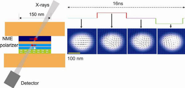
Figure 1.
Left: Schematic of the pillar structure, showing
the ferromagnetic layers in blue, the antiferromagnetic
pinning layer in green and the Cu leads and spacer
layers in orange. The bottom two FM layers are coupled
into a fixed antiferromagnetic arrangement by a Ru
spacer layer and their magnetization direction is pinned
by exchange coupling to the green antiferromagnet shown
at the bottom. The incident X-ray beam is incident 300
from the surface normal and is focused by a zone plate
to a size of about 30 nm. The transmission through the
structure as a function of sample position is monitored
by an X-ray detector.
Right: Measured time dependent magnetization
directions indicated by arrows within the 4 nm
thick Co0.86Fe0.14
sensor layer inside the 100 nm x 150 nm nanopillar.
Images at four times are shown. By varying the delay
time of the X-ray probe pulse relative to the current
pulse entire motion pictures were recorded.
Motion pictures with 200 picosecond time resolution and
30 nm spatial resolution reveal a fast, subnanosecond
switching process based on the lateral displacement of a
magnetic vortex, as illustrated at four selected times
in Figure 1. While the motion of vortices is omnipresent
in nature their role in magnetic switching has
previously remained unrecognized or unappreciated. Our
measurements show how the injected spin current
laterally displaces magnetic vortices created by the
curled Oersted field of the accompanying charge current.
The new fundamental switching process is intriguing in
that it is accelerated by the curly Oersted field yet it
may result in metastable final states which are
undesirable in technological applications.
Now that the experiment technique has been established
and demonstrated different magnetic configurations and
sample geometries will be explored. Of particular
interest are sample geometries where the electrical part
of the current does not directly flow through the sensor
layer but the effect of only the spin polarized
electrons that diffuse into the sensor layer can be
studied. This will be explored with new types of samples
which do not have the form of a pillar but are planar.
One of the main questions is whether the switching time
in such “spin-current only structures” will be decreased
by the absence of the Oersted field which accompanies
the charge current.
8. Behavior of Charges, Excitons and Plasmons at
Organic/Inorganic Interfaces
M.D. McGehee, N. Melosh, M. Brongersma
As electronic device dimensions shrink to nanometer
scales and the range of desirable applications grows,
two trends are emerging. First, the range of materials
under serious development is growing, and many device
structures consist of both organic and inorganic
building blocks. Second, many physical phenomena that
were heretofore only observed within academic
experiments are becoming important for technologically
relevant devices. Consequently, there are a large number
of technical issues that need to be solved before these
new possibilities become technologically viable. These
include reproducible device performance on this length
scale, sample heterogeneity, interface state control,
defect properties, thermal transport and surface
roughness. In addition, physical phenomena such as
electron tunneling, Förster coupling, and
plasmon-excitation quenching begin to severely impact
device behavior at length scales less than 10 nm. This
is particularly true within the emerging subset of
structures that utilize both organic and inorganic
materials, such as solar cells, electronic paper,
molecular electronics, and organic light emitting
displays. Within this rather broad collection of
challenges, our team has identified the need to
understand excited state behavior within organic species
close to inorganic surfaces as a key problem for future
applications of these materials.
Excited state phenomena within organic materials are
often complicated by the multiple length scales,
morphology, multiple competing decay processes, and
inorganic surface interactions that affect the overall
behavior of the system. Current studies of realistic
devices are complicated by simultaneous excitation decay
via a number of different processes within different
regions of the sample. These decay processes, in which
the charge or energy of an excited state in a molecule
is transferred to an adjacent metal electrode, depend
strongly upon the molecule to metal spacing.
In order to address these issues systematically, our
team examines exciton transfer and decay within organic
systems on a hierarchy of length scales. Melosh studies
exciton transport and molecular wave function coupling
to metal electrodes on the molecular level (~1 nm).
McGehee examines exciton and charge transport within
conducting polymer films close to metallic electrodes or
dielectric films (5-100 nm). Brongersma investigates how
excitons couple to surface plasmon waves on metal
surfaces within the 10-500 nm range. Collectively, these
measurements provide a better overall understanding of
the behavior and importance of charges, excitons and
plasmons within electrically active organic-inorganics
than would be possible from a single study alone.
Charge Transport in Conjugated Polymers:
The structure has been characterized and the charge
carrier mobility measured of films of regioregular poly
(3-hexylthiophene) P3HT as a function ofmolecular
weight, casting solvent, annealing temperature and
surface treatment. The primary characterization
technique was X-ray diffraction at SSRL. It has been
seen that when low molecular weight polymers and low
boiling point solvents are used, crystals form rapidly
in the bulk of the film. Consequently the crystals do
not align with each other and the conjugated parts of the
polymer chains do not stack up against each other at the
grain boundaries. The charge carrier mobility can be as
low as 10-6
cm2/Vs.
On the other hand, when higher molecular weight polymer
and higher boiling point solvents are used,
crystallization tends to occur only at the surface of
the gate dielectric. Consequently, the crystals all tend
to have the same orientation and charge carriers can
easily hop from one crystal to another. The mobility in
this case can be as high as 10-1
cm2/Vs.These
studies highlight how critical it is to optimize the
structure of organic semiconductor films when assessing
new molecules for applications. The initial
demonstration used P3HT as the light absorbing and
energy donating material. This polymer is actually a
poor choice because it is not a good light emitter and
therefore is not a good energy donor by the Förster
mechanism. Better emitters have been used and it was
found that energy transfer becomes effective over
distances as large as 30 nm. Unfortunately, the energy
levels of the polymer used were not suitable for
splitting excitons at the interface between the two
polymers. A goal for this year is to obtain suitable
polymers from the Molecular Foundry and Jean Fréchet’s
group at UC Berkeley so that solar cells that utilize
energy transfer over 30 nm distances can be made.
Exciton Transport in Conjugated Polymers:
Almost all organic photovoltaic cells are based on
either planar or bulk heterojunctions of two
semiconductors. After light is absorbed, excitons must
get to the interface between the two semiconductors to
dissociate by electron transfer. In some cases, such as
in dye-sensitized cells or polymer-fullerene bulk
heterojunctions with very high fullerene concentrations,
excitons are formed right at the interface and exciton
transport is therefore not a limiting factor on the
performance of the cells. In many other cases, such as
in polymer-nanowire or polymer-titania cells, excitons
need to travel at least five nanometers, if not more.
For this reason exciton diffusion is a very important
process to understand and optimize. Exciton diffusion
must also be avoided in light-emitting diodes so that
excitons do not reach quenching sites. The exciton
diffusion length has been measured in several polymers
and it was found that the values are less than reported
in the literature. Common sources of error in diffusion
length measurements are neglecting interdiffusion
between the donor and acceptor, interference effects and
resonance energy transfer. Since the diffusion length in
most polymers is 6 nm or less, ways have been explored
to enhance exciton transport. One is to use resonance
energy transfer from a donor to an acceptor with a
slightly smaller energy gap. It has been shown that
exciton harvesting form P3HT, a polymer commonly used in
the best polymer solar cells, can be improved by a
factor of three by transferring energy to a low band gap
polymer. This improvement in exciton harvesting triples
the efficiency of solar cells made with P3HT. It has
also been shown that resonance energy transfer occurs in
many previously studied donor-acceptor blends, including
polymer-fullerene blends with low fullerene
concentrations.
Determining the Orientation of Organic Molecules at
Buried Interfaces: It has recently beenshown that crystals nucleate at the
interface with the dielectric in polymer thin film
transistors by analyzing XRD rocking curves and
observing that the crystals could only be as well
oriented as they are if they nucleate off of the
interface. This technique will be used to address the
general
question of how conjugated polymers pack at interfaces.
This topic is particularly relevant to organic-inorganic
solar cells. It is hypothesized that in some cases
polymers, such as P3HT, pack with their side chains
pointing towards a titania surface. For reasons not yet
understood, in other cases the polymer packs with its
conjugated backbone attached to the titania. It is
thought that the cells with the backbone attached to the
titania work much better because there is no barrier to
electron transfer.
Omni-Directional Emitters and Plasmon Coupling: Brongersma demonstrated in a series of experiments and
electromagnetic simulations that metal/light emitting
molecule/metal optical microcavities can be engineered
to produce an omnidirectional emission resonance. The
two metal layers in this cavity have a clear optical
function, but at the same time they also serve as
electrical contacts for current injection. This is quite
desirable from an electronics viewpoint as metals
exhibit a low resistance. However, in current light
emitting devices metals are generally avoided and more
resistive transparent conductors such as Indium Tin
Oxide are used as they allow for more efficient light
extraction. Brongersma is interested in further
exploring the use of metals in light emitters and
exploiting the unique properties of surface
plasmon-polaritons to increase the out-coupling
efficiency, decrease the lifetime of emitters, and
modify the angular emission distribution. In this study,
he showed that for a cavity thickness equal to
one-quarter of the surface
plasmon resonance wavelength,
λsp/4,
a completely flat dispersion relation for surface
plasmonpolaritons can be realized. Dipolar emitters
placed in such a cavity radiatively decay into surface
plasmon-polariton modes that subsequently couple to
far-field radiation in all directions. This
omni-directional resonance contrasts sharply with the
typically highly directional resonant enhancements from
waveguided modes in planar dielectric optical
microcavities. In order to experimentally verify the
predicted behavior, the angular emission from a
blue-emitting polymer within a gold cavity was measured.
In these experiments the omni-directional behavior was
indeed observed and the work is currently being
submitted to Applied Physics Letters. These studies may
be of importance for the field of solid state light
emission, which is rapidly gaining in importance in a
number of applications such as traffic lights, room
lighting, and displays. In particular, the observed
isotropic emission is important in applications where a
large viewing angle is desired.
Surface Plasmon Spectroscopy:
The Melosh group has focused on investigating the
optical properties of molecular films within
metal-molecule-metal junctions and their behavior during
electrical cycling. The goal is to resolve the dispute
about molecular re-arrangements under high bias that may
lead to different molecular conductivity, which has
special importance for molecular electronics and
understanding electron transfer near metal electrodes. A
new technique has been developed, ‘Surface plasmon
spectroscopy’
(SPS) to probe the optical absorption of molecular
layers between metal electrodes. This method enables us
to ‘see’ inside a 1-3 nm thick molecular film between
two opaque metal electrodes. Based upon the Kretschman
surface plasmon excitation architecture, this method
relies upon the reflectivity change due to coupling into
surface plasmons to measure the real and imaginary parts
of the molecular film’s dielectric constant. By
repeating this measurement at multiple wavelengths the
optical absorption spectrum can be obtained from the
imaginary component of the dielectric constant.
It was discovered that the absorption of molecules on a
single
metal surface is almost identical to solution phase,
however thin organic films placed between metal
electrodes exhibit significant absorption maxima shifts
(15-30 nm). These shifts are believed to be due to Stark
effects within the junctions, and need to be included in
electron transport calculations. Typically these
junctions consist of an Au or Al bottom electrode 20-30
nm thick, a thin molecular layer 5-30 nm thick, anda top
Au metal contact 15-30 nm thick. However, to
simultaneously test the electrical response and optical
absorptivity, new top metal contact methods are
necessary to prevent pin-hole shorts through the
molecular layer.
The limits of Surface Plasmon Spectroscopy will be
pushed further in order to determine its maximum
sensitivity and resolution. Measurements will be
extended to include single molecular thick films of dye
molecules R6G and ‘active’ molecules such as spiropyrans
and [2]-rotaxanes. These active molecules change their
optical, and presumably electronic, state upon either
UV absorption or electrical redox chemistry,
respectively. Studying the molecular reconfigurations
using SPS within these systems will help us understand
the nature of molecular electronic behavior in direct
contact with electrodes, an important topic for emerging
applications utilizing organic species together with
inorganic structures.
In addition to direct optical studies, the change in
optical properties will be examined as electrical bias is
applied to metal-molecule-metal junctions. Molecules may
include model systems such as alkanes, or conjugated
systems like oligo-phenylethylene vinylene (OPEs). In
order to take full advantage of these molecules, the
PALO technique must be extended to be able to deposit
the top float-on electrode onto a Au bottom electrode,
which currently results in shorting. We believe this is
due to the high mobility of the Au atoms under large
applied bias, thus we are going to investigate methods
to lift on Al top electrodes onto Au bottom electrodes.
Surface Plasmon Sources: Brongersma will continue his research in the area of
Metal-Molecule-Metal junctions. In addition to using
these junctions as light sources, he is interested in
exploring the possibility to fabricate a surface
plasmon-polariton source. To this end, new simulation
tools will be developed that can quantify the emission
out of the side of the junction and into surface
plasmon-polariton modes. The Metal-Molecule-Metal
junctions will be attached to a metal (plasmonic)
waveguide that can guide the surface plasmon-polaritons
over short (~ 10 μm)distances. Interestingly, the metal
waveguide can again serve a dual purpose and act as the
electrical contact to the source as well. The end goal
is to fabricate a stable, low-noise, high intensity, and
compact plasmonic source. These type of pigtailed
sources could be of practical importance and can be used
to ensure efficient coupling from light sources to
optical waveguides and fibers.
Soft Contact Metal Deposition:
In order to address electrical shorting in our
evaporated metal devices, a new technique was developed
to softly deposit the top metal contacts onto an organic
film. Depositing the top metal contact for molecular
electronics is always difficult, especially for
near-atomically flat films. Sputter or e-beam deposited
metals always damage the organic sample to some extent,
while other ‘soft’ methods like lift-off float-on
electrodes wrinkle excessively and stamped contacts are
atomically quite rough. Our procedure, “Polymer Assisted
Lift Off” (PALO), utilized a thin polymer as a backing
to support pre-evaporated metal electrodes, which gave them mechanical strength and hydrophobicity. Using a
nonadhesive metal deposited onto an ultra-flat substrate
(mica or Si), the electrodes could be ‘lifted-off’ onto
a water surface due to the hydrophobicity of the polymer
(e.g, PMMA) layer. This floating set of electrodes could then
be transferred onto a thin organic film without damage. A
paper on the PALO method is under review by
Advanced Materials.
Large electrode arrays were created with the PALO
technique, with wire widths as small as 1 micron to as
large as 3 mm, with lengths up to 3 cm. This is an
extraordinary dimension for 20 nm thick metal films, (an
aspect ration of 1.5 million!) especially without any
wrinkling or buckling of the metal. This is a result of
the thermodynamic driving force of the polymer layer to
force the water from between the two surfaces, and the
favorable maximization of the metal –water surface area.
To demonstrate the utility of this technique, we
performed studies of electron tunneling through carboxy
alkanes with a PALO top contact. These devices
demonstrated the same electron tunneling behavior as
literature STM and nanopore measurements. Even with 4 mm2
contact areas, no shorting is observed.
9. Development and Mechanistic
Characterization of Alloy Fuel Cell Catalysts
A. Nilsson, P. Strasser, H. Ogasawara
The main focus of this research program is the
investigation of mechanistic aspects of fuel cell
catalysis on metal surfaces. One of the main
challenges for the Hydrogen Fuel Initiative is to
develop cost efficient electrocatalysts with high
durability for the next generation of fuel cells. An
essential aspect of this project is to develop
synchrotron radiation based X-ray diffraction and
spectroscopy methods that allow
in-situ
probing of the intermediates in the catalytic
cathode process where both species identification,
geometric and electronic structure properties is
fully characterized. In parallel to the fundamental
synchrotron work, theory-guided combinatorial
synthesis and high throughput electrochemical
screening methodologies for fuel cell cathode
catalysts will be developed and applied in order to
link mechanistic hypotheses and catalyst testing
under realistic conditions in high dimensional
compositional and process parameter spaces.
Oxygen Reduction by Water on Surfaces:
The adsorption of water on oxygen covered metal
surfaces reduces oxygen to OH forming a OH-water
co-adsorbed phase. This OH-water coadsorbed phase is
an important intermediate in fuel cell catalysis.
XAS, XPS and STM studies were performed on the
OH-water co-adsorbed phase on Pt(111) and Ru(001)
surfaces. While the OH species is fully hydrogen
bonded to surrounding water molecules on Pt(111),
the OH species on Ru(001) is in a non-donor broken
hydrogen bonded configuration.
Wetting at Water on Surfaces:
The fuel cell reaction occurs in confined spatial
regions called triple phase boundaries where the
gas, electrolyte containing water and catalytic
metal particle contact. A wetting behavior of metal
surface plays an important role in how triple phase
boundaries. We demonstrated on Pt and Cu surfaces
that the wetting is related to the difference in
substrate electronic structure.
Instrument Development:
An electron spectrometer equipped with three
differential pumping stages was installed on surface
science end-station at SSRL BL5-1 for the
in-situ
investigation offuel cell reaction. A differentially
pumped ambient-pressure reaction cell using
cryogenic technology is being assembled.
Structural Molecular Biology
The primary purpose of work described here is to develop
synchrotron radiation facilities and provide access for
the national scientific community through a strong user
support program. Such synchrotron resources are a
powerful and versatile tool for research in structural
molecular biology, and provide tools very relevant to
addressing the U.S. Department of Energy mission needs.
The scientific and technological focus of this program
includes the applications of synchrotron radiation to
macromolecular crystallography, small-angle X-ray
scattering (SAXS) and X-ray absorption spectroscopy
(XAS). These efforts are led at SSRL by Professors K.O.
Hodgson, B. Hedman and W.I. Weis, and Drs. S.M. Soltis
and H. Tsuruta.
Key aspects of the program being provided by the BER
funding include:
-
Continued availability to, and support of users on,
state-of-the-art beam lines and instrumentation on
the upgraded 3rd-generation
SPEAR3 storage ring for SMB research for a
significant fraction of a given year (~9 months or
more per year)).
-
Enhanced user support and training for SMB
scientists using up to 10 existing stations at SSRL
(of which eight are on high-intensity, multipole
wiggler beam lines).
-
Full operation and user research program on all
three stations on the Beam Line 9 facility dedicated
to SMB research.
-
Continued development and implementation of advanced
optics, experimental facilities, detectors, computer
resources and software to enable optimal advantage
to SMB users of the capabilities of the new 3rd
generation SPEAR3 storage ring.
-
Continuation of capital improvement projects in
areas such as beam line enhancements, data
acquisition systems including detectors,
electronics, controls and computer hardware for SMB
stations.
-
Continued synergistic research and user support in
the SMB area with the NIH National Center for
Research Resources (NCRR)-funded Biomedical
Technology Program (BTP) and the National Institute
of General Medical Sciences (NIGMS)-funded
macromolecular crystallography operations support
and Structure Determination Core of the Joint Center
for Structural Genomics.
BER Funded Staffing and New Opportunities for SMB R&D
The BER-funded scientific and technical staff at SSRL
currently effectively support users of up to ten
existing stations, including the three BER-funded Beam
Line 9 SMB stations (BL9-1 and BL92 for macromolecular
crystallography and BL9-3 for X-ray absorption
spectroscopy). This is done in coordination with other
specialized activities supported by NIH NCRR and NIH
NIGMS. Ph.D.level research staff, currently all or in
part supported by BER, are A. Cohen, R.P. Phizackerley,
C. Smith, S.M. Soltis (in crystallography), S. DeBeer
George, B. Hedman, K.O. Hodgson (in X-ray absorption
spectroscopy), and H. Tsuruta (in small-angle X-ray
scattering). Support is also continued for one summer
month of salary support for Professor W.I. Weis (a term
member of the SSRL faculty with primary appointment in
the Department of Structural Biology on Stanford campus;
a leading expert in multiple-wavelength anomalous
dispersion (MAD) phasing who contributes significantly
to continued developments and applications in this
important area).
Professor A. Brünger has a joint appointment between
SSRL (⅓) and the Stanford School of Medicine (⅔). His
activities focus on computational and methodological
macromolecular crystallography, and are having a very
positive impact on providing new capabilities for SSRL
users and staff. Funding continued in FY2005 and FY2006
for support of a graduate student research assistant (S.
Kaiser) to work with Dr. Brünger on SSRL SMB-related
developments.
Five-Year SMB Program Plan for Beam Line and
Instrumentation Developments and for User Operations
Support
– A competitive five-year renewal proposal for the
DOE-BER and NIH-NCRR funded SMB Resource at SSRL was
submitted formally to NIH on June 1, 2004. As per
discussion and agreement between the DOE-BER and
NIH-NCRR program staff, this renewal formed the basis
for a joint evaluation of the synergistically funded and
managed SMB program at SSRL. The proposal contained
developments directed specifically to the BER-funded
program, which focuses mainly on developments and
implementation of new instrumentation and beam line
facilities to enable the SMB user community to benefit
in the most optimum way from the new capabilities
provided by the new SPEAR3 accelerator. The Special
Study Section Committee, which performed a site visit at
SSRL in November 2004, provided an exceptionally strong
endorsement of the SSRL SMB program as a whole, and
recommended that the BER funds requested for personnel,
travel, materials and supplies and other non-equipment
expenses, be provided at the budget level requested in
the proposal. For equipment, it was recommended that all
proposed items in the BER budget be funded with the one
modification: a reduction in scope of a data storage
system, from 300 TB to 100 TB. A FWP, modified according
to the Study Section Committee recommendations and with
a revised budget, was submitted to DOE-BER on 4/25/05.
The proposed Operations Funding and Capital Equipment
Funding in this FWP have since been revised and is
consistent with programmatic guidance. The NIH-NCRR
grant award for the five-year period of the SSRL SMB
program has been made.
Structural Molecular Biology Program at SSRL
During the FY2006 run, the SPEAR3 accelerator continued
to be exceptionally reliable, providing very stable beam
for a very high fraction (96.2%) of the scheduled time,
at up to 60+ hr life times. The new rapid fills, taking
1-3 mins each, became routine and greatly enhanced the
use of beamtime and the thermal stability of the optics.
The FY2006 user run extended over a ~8 month period from
~November 28 through August 7, 2065. Outstanding
utilization and performance were seen over the run
period. About 50% of the proposals assigned time was for
structural molecular biology-related research. Demand
for beam time continues to be greater than the available
beamtime.
SSRL is planning a normal ~9 month/year run cycle,
delivering (as do the other three DOE synchrotrons)
about 5,000 user hours per year (i.e.,
about 630 scheduled user shifts per year).SSRL has the
goal of maintaining or increasing this by as much as 10%
over the coming 3-5 years, paced by the needs for
operational shutdowns for new beam line insertion
devices and other planned upgrades and the availability
of adequate base facility operating budgets from DOE BES
(which are anticipated given the budget outlook for
DOE-SC in the coming 10 years).
The strong demand for all the stations at SSRL
continues. Over all the stations at SSRL, the overdemand
averaged ~160%. Among the most severe in the overdemand
category remain the crystallography stations, with a
combined overdemand of ~230% for beam lines BL9-1, BL9-2
and BL11-1. The SAXS wiggler station BL4-2 was in
overdemand by 176% despite having become a dedicated
beam line for this technique and science, whereas BL9-3
(for XAS) was in overdemand by 267%, one of the highest
in all of SSRL. The fraction of beam time on X-ray
stations allocated to structural molecular biology
research continues to be ~35-40% while the fraction of
SMB users is ~45%. Since the majority of this beam time
is awarded on a peer-review competitive basis among all
SSRL proposals, SMB proposals continue to compete very
well at SSRL.
User Satisfaction
– As continuing part of user activities at SSRL, each
user group is asked at the end of their run to complete
an "End-of-run summary form". This form provides an
opportunity for pointing out specific problems/issues
and offering suggestions as to means of improvement. It
also asks several questions to get a reading on overall
satisfaction. The forms are analyzed by the SSRL staff
and summaries by the SSRL Users Organization Executive
Committee. The users rate in five categories
(Unsatisfactory to Excellent). In the area of "Overall
Scientific Experience" 24% overall were very good and
68% were excellent in FY2005 the FY2006 statistics are
being compiled as of the writing of this document. These
summaries included all the open SMB beamlines and users
and, while subjective, indicate a significant measure of
user satisfaction with the operation and service of the
facility.
Beam Line 9 Upgrade Project
– At the end of the FY2005 run, there remained, to
replace the BL9-1 and BL9-2 monochromators as well as
about 80% of the beam line masks, slits, filters, and
windows. Additionally, motivated by the expected
improvement in beam focus, the BL9-2 branch line needed
to be realigned 0.4 mrad to the insertion device
centerline. At the beginning of the 2005 shutdown, the
initial focus was the installation, alignment, plumbing,
cabling, and motion control tests of the in-alcove
hardware. The BL9 in-alcove installation was completed
successfully by November 2005, when attention turned to
the out-of-alcove hardware installation. The BL9-1 and
BL9-2 monochromators and the rest of the branch line
hardware components for all three lines were installed
and alignment was completed by January 2006. The
low-conductivity water (LCW) system and associated
machine protection system (MPS) sensors were replaced.
Branch line commissioning began in late January.
Staggered starts of user operations of all three branch
lines (in order BLs 9-3, 9-2 and 9-1) took place during
the month of February, and they are now in full use by
the general user community. The upgraded optical
components have already led to a demonstrated increased
performance of 2X and 3X for BL9-1 and 9-2,
respectively, over earlier operating conditions with
SPEAR3. All branch lines for BL9 are now ready for
SPEAR3 500-mAoperations, which are anticipated to
commence during specific periods in the next year’s run
cycle.
Beam Line 4 Upgrade Project
– The BL4 500-mA upgrade project is yet to be completed
due to overall budget constraints. Despite the
comparative lack of resources (due mainly to the
constrained DOE BES core budget that is sharing the
upgraded cost of the BL and the significant
cut in those funds in FY2006), many BL4 components
including the LN2-cooled monochromators, the monochromator slits, the graphite
filters, the Be windows, and the branch line beam
stoppers are in various stages of fabrication and
assembly or are complete awaiting installation. In the
coming months the mirror systems, mirror slits, fan
allocation masks, pivot masks, and hutches will become
the major focus of the BL4 upgrade effort.
One major and unexpected development in the BL4 upgrade
project was the decision in FY2005 to relocate BL4 from
its current location in Building 131 to adjacent to BL11
in Building 130.
This decision rests on several factors: (a) Rebuilding
BL4 in a new location permits installation activities to
commence while BL4-2 continues to serve the Bio-SAXS
user community. This will minimize the BL4 down time
since the existing BL4 need not be decommissioned until
much of the new BL4 is already installed. (The relocated
and upgraded BL4 will reuse only the existing BL4
insertion device, front end, hutch instrumentation, and
much of the instrumentation and control suite.) (b) The
new BL4 location incorporates a portion of the SPEAR3
concrete shield wall that already has been upgraded to
current seismic standards. In contrast, the SPEAR3
concrete shield wall at the old BL4 location was
scheduled for seismic retrofit in several years. Since
the retrofit of this wall necessitates removal of nearby
beam line hardware, relocation ofBL4 avoids future
disruptions of the beam line during this seismic
retrofit. (c) The relatively unencumbered geometry of
the new BL4 location simplifies the installation while
affording the opportunity to optimize the beam line
layout for improved performance, functionality, and user
ergonomics. BL4-2 will continue in its new location to
serve at 100% the general user community in the area of
biological small-angle X-ray scattering/diffraction.
Installation and recommissioning of the new BL is
anticipated to be complete in FY2007.
Beam Line 7 Upgrade Project
– The end of FY2005 and the beginning of 2006 saw a
flurry of activity and associated progress on the BL7
500-mA upgrade project such that the beam line at this
point is a few working weeks away from the start of
commissioning. Much of early FY2005 was spent assembling
the remaining BL7 masks and optical systems in
preparation for installation. The masks and mirrors to
be installed inside the SPEAR3 shielding enclosure
received the highest priority for attention to ensure
that all such components were ready for installation
during the tight summer 2005 installation envelope. The
optical configuration of each branch line employs an in-alcove
M0
mirror for power filtering, harmonic rejection, and beam
vertical focusing (BL7-1) or beam collimation (BL7-3).
These single crystal silicon mirrors are side cooled
through contact to water-cooled copper pads using a
gallium-indium eutectic as a thermal transfer medium.
During this same time period, the remaining
“out-of-alcove” optical elements, masks, slits, windows,
and
vacuum transport systems were assembled. Several key
elements, such as the BL7-3 LN2-cooled monochromator, were assembled in prior years and stored
for later installation. Among the key components
assembled in FY2005 was the BL7-1 side scattering
monochromator. This focusing monochromator employs a
side-cooled, cube-root cut Si crystal assembly, like
BL9-1 and BL11-1.
By the start of the SPEAR3 summer shutdown in August the
vacuum assembly of the beam line optics and beam
transport components was largely complete and the effort
turned to the rapid disassembly and removal of the
obsolete BL7 components and radiation enclosures
(hutches). Within approximately one month from the start
of the shutdown, the site of BL7 was stripped bare of
virtually all optics, support hardware, and hutches, and
this removal of the old hardware paved the way for
installation of the new beam line. The most important
installation milestone was the completion of the
installation, alignment, plumbing, cabling, and motion
control tests of the in-alcove optics and beam transport
hardware prior to the close up and restart of SPEAR3 at
the beginning of November 2006. While the in-alcove
components were being installed, new radiation
containment hutches were constructed in the
out-of-alcove area. This task is nearing completion
atthis point, and the installation of the out-of-alcove
beam line optics and beam transport is near completion.
When complete, this rebuild will provide 100% general
user access to BL7-1 for macromolecular crystallography,
with the same infrastructure as provided for all other
MC beamlines at SSRL, and BL7-3 will be dedicated to
dilute solution biological X-ray absorption spectroscopy
(100% general user), alleviating some of the
oversubscription of BL9-3 and thereby providing more
time for other techniques, such as microXAS imaging and
single crystal XAS on BL9-3. The upgrade of BL’s 7-1 and
7-3 is mainly funded from NIH NCRR.
Macromolecular Crystallography
For macromolecular crystallography, advances in
instrumentation and beam line technologies continued in
FY2006. During the FY2006 run, five MC beam lines were
in operation with the new SPEAR3 lattice running at a
current of 100 mA (BL11-1, BL9-2, BL9-1, BL1-5 and 50%
of the available time on the shared station, BL11-3).
The Stanford Auto-Mounting (SAM) system was available on
these beam lines. The SAM system incorporates a robot
that can mount up to 288 samples without the user having
to enter the experimental hutch and operates in an
integrated environment within the Blu-Ice experiment
control software that can be used to select and screen
samples in a totally automated fashion. During FY2006,
the SAM system was used routinely by a significant
percentage of the user community. A high priority will
be placed on optimizing the beam line in-hutch
instrumentation for 500-mA SPEAR3 performance.
Specifically, the newly rebuilt BL7-1 is being
commissioned and made operational for the general user
community (and as mentioned – this phase took place for
BL9-1 and BL9-2 early in FY2006).
Beam Line 12 – New Capacity and New Experiments
– The California Institute of Technology received a
$12.7M gift from the Gordon and Betty Moore Foundation
to fund the construction of a new high-intensity,
state-of-the-art synchrotron beam line for MC research
at SSRL. Under a cooperative research agreement, the use
of the beam line will be shared between macromolecular
crystallography general user community (60%) and
scientists at the California Institute of Technology
(40%). BL12 will make use of a powerful in-vacuum hybrid
undulator source. The 67-period undulator with a 6 mm
gap operating on SPEAR3 will produce a brightness of ~1018
p/s/mrad2/mm2/0.1%bp
at an energy of 12 keV, approaching that of the APS UA
undulator. The beam line will provide a very narrow
energy band pass for optimized MAD experiments and is
being designed to accommodate the study of
microcrystals. With this very bright source, it is
projected that samples with dimensions on the order of
~5-10 µm on an edge, and otherwise weakly diffracting
crystals, will be able to be routinely studied.
The design and construction of BL12 is progressing well,
with commissioning currently scheduled for early FY2007.
The undulator ID was delivered in late June and has been
installed in SPEAR3. The four dipole magnets required to
produce the SPEAR3 orbit chicane for the new ID and the
associated new SPEAR3 vacuum chamber have been
installed. The quadrupole magnet triplet required to
focus the SPEAR3 beam into a double waist for the BL12
ID was fabricated and installed during the summer 2005
SPEAR3 shutdown. The new SPEAR3 magnetic lattice
incorporating the double waist was commissioned at 100
mA during the fall. More recently this lattice was
successfully injected and run at 500 mA. The beam line
front end has been installed. The three mirrors required
for the beam line optical concept are currently being
installed. The
LN2-cooled
monochromator hardware has been installed and testing
will commence shortly. Modifications to Building 131 and
the SPEAR3 concrete shielding enclosure required to
extract the beam were initiated and completed during the
summer 2005 SPEAR3 shutdown. Overall, the beam transport
systems are on schedule for completion by November 2006.
The hutch instrumentation is conceptually established,
with the standard SSRL crystallography equipment and
software being utilized wherever possible. The crystal
mounting robot, cryostreamand fluorescence detector
systems will be replicated with some improvements.
Modifications to the sample table and detector
positioner to increase the stability of the sample
handling system are being developed. The designs for
systems needed for the visualization and manipulation of
micro-samples have also been developed and are based on
currently available technologies. A Mar325 CCD detector
has been ordered for the beam line. BL12 will provide a
state-of-the-art facility for SSRL’s general users to
carry out extremely rapid data collection and to
routinely study challenging systems such as large
complexes, viruses, micro-crystals, and weakly
diffracting samples with large unit cells. Funding for
general user operation for this beam line was included
in the submission (and recommended) of the five-year
peer-reviewed proposal to DOE BER, but has not yet been
realized. It is our hope that funding for this purpose
can be added beyond inflationary increases in the
operations budget in FY2007.
Remote Data Collection – Since June 2005, macromolecular crystallography users
have had the option to conduct remote user access data
collection diffraction experiments from their home
institutions and other remote locations by means of
advanced software tools that enable control of the beam
lines. Remote experimenters have access to the same
tools as local users, and have the capability to mount,
center, and screen crystalline samples, and to collect,
analyze, and backup diffraction data. Automated sample
mounting is accomplished with the SAM system. Beam line
and experimental control is carried out using
Blu-Ice/DCS and additional remote monitoring of the
experiment and data backup is supported with several
web-based applications. The highly graphical
applications and computational resources at SSRL are
accessed through a client/server application that uses
minimal resources on the client side and has a typical
response close to that obtained at the beam line. This
remote capability is now available on beam lines 1-5,
9-1, 9-2, 111, and 11-3, and starting in FY2006, 7-1.
Many of the applications required to perform a
crystallography experiment are highly graphical in
nature, and typically do not perform well through
standard remote login techniques over large geographical
distances. This problem has been addressed with the use
of a terminal server application provided by NoMachine.
This server is accessible to remote users through a free
client application that is easily installed on their
home computers. This system enables the user to run all
command line and X-Window based applications available
at the beam lines, including the Blu-Ice control
software and data processing suites. SAM cassettes with
preloaded samples are shipped to SSRL and a staff member
loads up to three cassettes into the dispensing dewar at
the assigned beam line and authorizes the user group to
access the control system in a secure mode. Through a NX
client remote desktop, the experimenter selects and
mounts the samples from inside a cassette using the
Blu-Ice software to access the SAM system. After a
sample is mounted, it may be centered in the X-ray beam
automatically, or if desired, the experimenter may
manually adjust thecrystal position by clicking on the
video image, displayed at the home institution, of the
sample within Blu-Ice. The SAM system can be configured
for fully automated operation to screen large numbers of
crystals while unattended. From within Blu-Ice, multiple
samples can be selected for screening, and parameters
for acquiring test diffraction data can be configured.
An automated screening sequence is typically comprised
of mounting the sample, aligning the sample loop to the
X-ray beam, taking a diffraction image and video image
at a phi angle of 0 and 90 degrees, and returning the
sample into the cassette. This sequence is repeated for
all selected samples. Automated scoring of the
diffraction quality of each sample can be carried out.
The scoring results are written to a spreadsheet to
assist the user in selecting the best sample for data
collection. The user can then start data collection on
the best crystal, or a series of ranked crystals.
Users are becoming increasingly interested in operating
the beam line and accessing computational resources
remotely. Out of 65 beam time requests that were
received during the June and July scheduling period,
there were 18 requests (27%) for partial or full remote
access. For the first FY2006 scheduling period, 25
requests (46%) for remote access have been received. To
date, remote access has been used for more than 35
experimental user runs representing 20 research groups
as far away as Australia and New Zealand. During the
commissioning phase, NIAID members on a site visit at
The Scripps Research Institute in La Jolla observed the
remote screening of ~150 SARS related protein crystals
on BL11-1. This screening run aided in the structure
determination of a conserved domain from the severe
acute respiratory syndrome coronavirus [K.S. Saikatendu,
et al.,
Structure,
13,
1665 (2005)].
Other improvements to the macromolecular beam lines and
equipment include:
Automated Crystal Annealing
– Using automated crystal annealing techniques has been
shown to decrease mosaicity and improve diffraction for
some macromolecular crystal systems. SSRL now offers two
methods for users (on-site and remote) to anneal
samples, implemented through the Blu-Ice software and
GUI. During data collection, a cold nitrogen gas stream
flows over the crystalline sample to maintain it at
cryogenic temperature. The crystal is annealed by
letting the sample warm up to near room temperature, and
then quickly cooling the sample back down to ~100 K.
This may be initiated from the Blu-Ice GUI by either of
two methods:
stream blocking
or
flow control.
The annealing time for both methods is set by the user
and requires confirmation to prevent accidental
annealing. The
stream blocking
method uses a thin paddle to physically block the
nitrogen gas stream near the sample position. This
device may be controlled through the Blu-Ice GUI or
manually by pushing a button located on top of the unit.
The paddle position is encoded so that the control
system may check if the paddle is fully retracted out of
the gas stream. The device is also spring-loaded so that
the paddle will retract if power to the device is lost.
The main advantage of the
stream blocking
method is that the nitrogen stream is quickly blocked
and unblocked, rapidly warming and cooling the sample.
The second method for annealing,
flow control,
uses the nitrogen flow control settings of the beam line
Oxford Cryojet to turn off the nitrogen stream from
within the Cryojet dewar. Because there is a long
transfer line between the Cryojet dewar and the sample
position, the flow rate of the nitrogen stream at the
sample position is changed more gradually than with the
stream blocking
method. The main advantage of the
flow control
method is that only the cold nitrogen stream is stopped
and a warm nitrogen stream remains protecting the sample
from icing.
A Universal Sample Container for Robots
– The availability of robotic sample mounting systems
continues to increase at synchrotron sources. Most of
these are compatible with standard Hampton-style
magnetic cryo-pins. However, many types of containers
are used for sample pin transportation and storage.
Currently at synchrotron beam lines in the US there are
three main sample storage containers used: the SSRL
cassette, the ALS puck, and the Molecular Structure
Corporation (MSC) magazine. In an effort to minimize
compatibility problems for users, a collaboration was
initiated by SSRL with the ALS, the APS SBC-CAT and the
industrial company, MSC to develop a “universal puck”.
The universal container resembles a standard ALS puck.
The key differences between the universal container and
the original ALS puck are that the universal container
incorporates features of the SSRL cassette and provides
the same clearance around the sides and bottom of the
sample pin as an MSC magazine. Both the universal
container enclosure and the base contain magnets and the
assembly will be held together by magnetic force. All
compatibility issues have been addressed, and the
approach is believed to provide complete transferability
between all three systems.
The enclosure piece of the universal container will be
used inside the SSRL SAM dispensing dewar. To adapt the
enclosure piece to the standard SAM setup an adaptor
cassette has been developed. Four universal containers
may be inserted into one adaptor cassette. The adaptor
is then inserted inside the SSRL robot dispensing dewar
using the cassette transport handle. Within the SSRL
dispensing dewar the three cassette locations will each
be capable of interchangeably holding an SSRL cassette
or an adaptor cassette containing four universal
containers. Once the development is complete, SSRL users
will have the option to use SSRL cassettes (with a 288
total capacity) or universal containers (with 192
samples) and the SAM system will be programmed to sort
samples between these two options.
Beam Line Simulator
– Funding has been secured from a combination of DOE BER, NIGMS and NIH NCRR to build a macromolecular
crystallography off-line facility for crystal screening
and beam line instrumentation and software development.
This facility will contain the electronics, computing,
software, and hardware required to mimic a typical SSRL
crystallography beam line (albeit with much lower flux).
A Rigaku MicroMax X-ray generator source that was
procured by the Joint Center for Structural Genomics
(JCSG) group with funding from NIGMS will be installed
on this facility. JCSG will also fund the robotic system
and both JCSG and SMB Core staff will participate in the
construction of the beam line simulator. A BER-funded
high-precision computer-controlled table is in
production. The control electronics and the final beam
definition system have been acquired with funding from
NIH NCRR, while NIGMS funds are used to procure a kappa
goniometer, Cryo-Jet Cryo-cooler, detector positioning
system and associated electronics. BER funding is also
providing the enclosure and the infrastructure. Benefits
of this off-line resource include: 1) an increase in the
rate of new developments without using precious
synchrotron beam time, 2) the availability of an
additional screening facility, 3) provision for an X-ray
source for screening when SPEAR3 is down and 4) use as a
training location where new staff and users can receive
hands-on instruction.
X-ray Absorption Spectroscopy
BL9-3, dedicated to general user biological XAS, accepts
2 mrad of the wiggler fan as a side station on the
16-pole 2-T hybrid wiggler BL9. BL9-3 provides extremely
high intensity over a
broad X-ray energy range, with focused beam from ~4 keV
to ~35 keV. The LN2-cooled
monochromator has two sets of Si(220) crystal pairs that
can be brought in and out of the beam without breaking
vacuum, providing a choice of two different azimuthal
orientations of the 220
planes with different glitch patterns, allowing a choice
for a given element/energy range. Its M1
optic (after the monochromator) is configured such that
the table does not move vertically during scanning,
providing excellent stability and thus data quality.
With SPEAR3, the focal spot is ~0.4 x 3 mm FWHM,
producing a measured flux of ~2.2x10¹²/100
mA at 9000 eV with 1x4 mm apertures. A factor of five
will be gained at 500 mA. With careful adjustment of the
collimating
M0
mirror, the beam line produces an energy resolution
close to the theoretical limit. It is a superb BioXAS
station and has enabled studies at µM concentration
levels, bringing the experimental capabilities closer to
physiological metal level in biological systems.
The upgrade for SPEAR3 500-mA operation was described
above. The major impact of this upgrade has been to
improve the beam line shielding and install beam line
masks, slits, filters, and windows for the higher heat
load. A significant additional improvement resulting
from this upgrade is that the new pivot mask and beam
line breakout in the SPEAR3 shield wall have made
it possible to operate the BL9-3 M0
mirror at lower incidence angle (thus higher energy
cutoff). The operating range was during the FY2004 and
FY2005 runs limited to ~5-23 keV. The new operating
range at lower mirror angle will allow focused
operations >30 keV. In the future, if user demand so
requires and with additional shielding, fully unfocused
operations will be possible with the upper energy cutoff
determined by the Si(220) monochromator crystal limit of
~45 keV. A second improvement is that the liquid
nitrogen feed lines for cooling the monochromator
crystals were re-designed to allow remote operation by
improving the routing of the lines. The previous lines
required entry into the optics hutch (closing all three
BL9 branch lines) to route the lines manually to avoid
catching on other BL components and causing damage. This
change will provide a significant improvement in
efficiency for user-requested monochromator crystal
changes.
In the 2004 submission, it was proposed and strongly
endorsed that the current capabilities for measurements
in the 2-5 keV range be moved from BL6-2, and that BL3-3
(a bending magnet beam line) be rebuilt for this
purpose. It has more recently been realized that rather
than upgrade the BL3-3 soft X-ray station in its current
location, this branch line could be incorporated as a
branch line on a new bending magnet line, BL14. This
would allow for a more optimal optics layout and hutch
setup, as the location provides for a “green-field”
approach, at what is estimated to be the same cost. The
optical concept for the station envisions a collimating
mirror followed by a double crystal monochromator and a
refocusing mirror. This beam line will be located in
Building 130 between the new location of BL4 and BL13.
Detailed design for this new beam line began FY2006.
Other development projects in FY2006 include:
Installation and Commissioning of a Hard X-ray
Fluorescence MicroXAS Imaging System
– A fluorescence microXAS imaging system, based on
Kirkpatrick-Baez (KB) mirror optics, was made available
to users on BL6-2 during FY2006. The KB optics were
purchased from Xradia Inc. The KB optics is mounted on
an optical rail inside a helium box. This He path
reduces air absorption and scattering of the primary
X-ray beam, providing higher flux and less signal
background. The setup contains a high-precision slit
aperture mounted downstream of the first, vertically
focusing KB mirror, which enables varying the
intercepted beam size of the optics. This allows trading
focused flux for spot size by probing different areas
and locations of the mirror surface. The commissioning
began with the characterization of the X-ray beam at the
location of the virtual source slit downstream of the KB
optics. It was found that the beam line focus at this
position was very close to the theoretical beam size
obtained by ray tracing calculations. Depending on the
size of the virtual source, a smallest focus size of
about (2 (horz) x 3 (vert)) μm2
with approximately 5x10⁸
photons/sec/100 mA was measured by scanning a 100 μm
diameter tungsten wire through the X-ray focus. The
largest focus size is about (15 (horz) x 11 (vert)) μm2,
which is limited by the size of the beam line focus at
the virtual source position.
Samples are mounted on a high-resolution x,y,z,theta
sample stage allowing for sub-micron step sizes using a
goniometer head with various sample mounting options for
different types of samples. The sample itself can be
observed by a high resolution long distance optical
microscope with manual adjustable zoom, which is
attached to a video camera/video server. A pellicle
coated with about 100 nm of aluminum allows observing
the sample in beam direction as the beam is scanned
across the sample surface. This optical microscope is
attached to a CCD camera/video server read-out and will
simultaneously enable to optically locate and monitor
certain areas ofinterest of the sample, which can then
be easily moved in and out of the focal position.
Currently the entire emitted fluorescence spectrum is
detected as a function of position on the sample by an
existing single element Si(Li) detector. In addition, a
high resolution X-ray camera was developed in FY2005.
This camera uses a very thin Gadox (P43) scintillator.
An optical lens creates a magnified image of the X-ray
beam on the scintillator onto a video camera with a
magnification of about 2.5:1. This camera is important
for characterizing the shape of the X-ray beam at
various locations in the X-ray hutch. This camera
improves and facilitates the optical alignment of the
entire beam line as well as of the fluorescence microXAS
imaging instrument. The K-B optic was funded by DOE BES
and additional equipment and partial staff effort is
provided from the SMB XAS program.
Instrumentation Development
path is enclosed in a helium atmosphere. The existing
setup achieves this by using flexible tubing which
connects to a Plexiglas slit box (which includes a
fluorescent screen for beam alignment) and then to the
sample box. The setup requires manual adjustment of the
slits and can only accommodate one sample at a time.
Since at these energies external calibrations are
necessary, frequent changes are required between the sample(s) of interest and the reference calibrant. This
requires that the user enters the experimental hutch,
changes the sample, and then allows the helium
atmosphere to re-equilibrate. Although the setup is
functional, significant improvements have been defined
and a design is underway that will enable higher quality
data, enhanced ease of use and more efficient beam time
utilization.
Motorized slits in a KF-40 flanged vacuum- and
helium-compatible configuration have been procured as a
replacement for the manual slit system. The slits will
be coupled to the beam line exit port and the sample box
space using vacuum-compatible bellows, which will reduce
potential helium leakage. Down-stream of the slits, a
6-way cross will be used to house a motorized linear
motion feed-through, which will have both a fluorescent
screen for sample alignment and reference calibrants for
external calibration. A view port on one side of the
cross will allow for viewing the beam size and position
using an Applied Scientific Instruments LC150 camera,
with a display at the work area. The opposite port will
house a photodiode for fluorescent measurements from the
reference calibrant. The design of the sample box space
is still in progress, and will include both a shutter
system (to minimize beam induce radiation damage) and a
sample cooling system. During this year sample cooling
options have been explored and tested, including a
contact-cooled sample block (to -90º C), a
Peltier-cooled setup, and a LHe flow cryostat with
internal photo-diode detectors. In addition,
improvements are being made to the existing detector
electronics. This modified setup will be used on BL6-2,
and will eventually be moved to BL14, when this beam
line has been built (see below).
XAS Instrument Control System Software and Computing
Developments
– The SSRL developedInstrument Control System (ICS)
forms the primary interface between control hardware and
data collection software. The ICS software provides a
consistent, convenient yet flexible interface to a large
variety of different types of beam line hardware. These
include, but are not limited to motors
(D.C. and stepper), real-time clocks, counters,
analog-to-digital converters and digital-to-analog
converters. ICS supports devices controlled using CAMAC,
VXI/VME, PXI, network, and RS232 based technologies. At
present ~70 different devices are supported. The ICS
software also includes a comprehensive set of
subroutines for client programs that provide easy-to-use
access to all devices in both a synchronous and
asynchronous manner. During FY2005 several improvements,
modifications and new instrument interfaces were added
to the ICS software. It should be noted that all such
changes have been made in such a way so that they can be
easily ported to the new ICS software (see below) with a
minimum effort. New device interfaces added during
FY2005 include: Lakeshore Temperature Controller, V535
VXI Stepper Motor Controller, Proteus XES Stepper Motor
Controller, Newport XPS D.C. Servo Controller, Ortec
Analog-to-Digital Converters, M550 CAMAC LVDT Reader,
and a Vortex & XIA DXP Detector Electronics Interface.
During the year, development was started on ICS
interfaces for both the Radiant Vortex-EX and XIA DXP
detector electronics. The Vortex software was used very
successfully on BL9-3, both by SSRL staff and visiting
scientists. It is anticipated that development of both
the interfaces will continue in FY2006 and beyond.
Although the current production version of ICS (V.1.22)
provides a robust, reliable and fully featured software
interface, it can only be used on Alpha CPU based
computers running the OpenVMS operating system.
Unfortunately, Alpha CPU based technology is in the
process of being phased out. A transition to different
platforms is being conducted in three main areas: (1)
the development of an Operating System Independent
version of the ICS software; (2) the migration of SSRL
Data Acquisition Software to the new Operating System
Independent model; and (3) new computer systems to
support legacy software during the transition away from
Alpha based CPU computers. Significant progress was
achieved in all three areas, and the new ICS software
was commissioned on BL7-3 in the spring of 2006.
s for XAS in the 2-5 keV Energy Region
–
An improved setup for XAS measurements in the 2-5 keV
range is currently in development, in preparation for
the new capabilities to be provided by a new bending
magnet beam line, as proposed in the program proposal.
In this energy range, X-rays have a very short path
length in air and thus the entire beam.
Also, significant progress was made in the design and
development of a dedicated beam line network.
Specifically a dedicated firewall machine was purchased,
a Fortinet Fortigate 1000 (with a combination of DOE-BER
and SMB DOE-BES funds). This firewall machine will be
installedas a “fail-over” partner to an existing
Fortigate 1000 firewall that already protects the SPEAR3
and Injector control systems. In addition, progress was
made on the design of the Virtual Local Area Networks,
or VLANs, that the firewall will provide. The design of
the network is being undertaken with the assistance of
the SLAC computer security team. It is anticipated that
BL7-3 will be the first beam line to take full advantage
of the new network architecture.
Small Angle X-ray Scattering/Diffraction
SAXS/D Beam Line 4-2 Operations
–
The SSRL small-angle X-ray scattering/diffraction
facility on BL4-2 is dedicated to small angle X-ray
scattering studies on biological systems. During the
FY2006 run, the excellent beam characteristics created
by the combination of the new low-emittance storage ring
and the 20-pole wiggler, provided users with enhanced
capabilities. Especially notable was the
high stability of the photon beam as a direct result of
the top-up injection scheme, which typically took 2-3
minutes three times a day, and helped stabilize the
overall performance of the optics components. The stored
electron ring current changed less than 20% between
fills at 60+ hr lifetimes and the practically constant
thermal load on the optics components resulted in the
high beam stability.
A number of user/staff research projects benefited
strongly from these beam characteristics. It is also
important to point out that several new exciting
scientific projects were also initiated during FY2005,
suggesting further growth in beam time demand and
continuing need for advanced instrumentation for
conducting challenging experiments.
SAXS/D Hardware Upgrades
–
In order to take advantage of the high-brightness SPEAR3
beam, a new instrument was built in FY2004. The main
goals of this development were to 1) extend the range of
instrument to both large characteristic length as well
as to higher structural resolution, and 2) allow
multiple instrument configurations during short beam
time by automation. The new instrument incorporates the
ability for automatically changing the
sample-to-detector distance and a pair of built-in
collimator/analyzer crystals in Bonse-Hart geometry for
ultra-small angle X-ray scattering (USAXS) studies. The
former feature allows users to select a desired
sample-to-detector distance, thereby angular range,
among five pre-designed distances, 0.5, 0.9, 1.4, 2.0,
and 2.5 m. The Bragg spacings, d=1/(2sinθ/λ), in Å
covered by these distance ranges are: 4.6-270, 8.7-520,
13-790, 18-1100, 23-1400, respectively, assuming the use
of a 9 keV X-ray beam and a 5-mm-diameter beam stop.
Development projects include:
Camera Improvements
– For the new camera, developments focused on automated
camera distance change and related improvements: (1)
Several hardware improvements were made to facilitate
distance changes. Three stages on a common optical rail,
individually supporting the most upstream flange of the
scattering path, the sample stage and the slit assembly,
were linked together in such a way that a single
motorized motion moves all at the same time, eliminating
manual translation of the sample stage. (2) A new
collimator assembly was built, consisting of two
in-vacuum slits built by Advanced Design Consulting, two
flexible bellows and vertical and horizontal translation
stages. The distance between the two slits is adjustable
between ~0.5 to 1.2 m, so that the up-stream slit can be
used as a beam defining slit for shorter
sample-to-detector distances. (3) The vertical
translation stage for the sample position was replaced
with one that provides a longer range. (4) Smaller
hardware improvements for the camera setup include the
design of a new static measurement cell holder, which
features dry nitrogen paths around each sample cell slot
to prevent moisture condensation so that measurements
can be performed at temperatures significantly lower
than room temperature,
e.g.,
4°C. The ability to simultaneously achieve
quasi-anaerobic conditions by nitrogen purging can be
useful for those samples that are sealed in the standard
sample cell, but normally require handling in a glove
box filled with nitrogen gas. (5) The sample alignment
video microscope was equipped with miniature vertical
and horizontal translation stages and the cross hair can
now be aligned with the direct beam position in the
Blu-Ice software sample monitor window so that the user
simply clicks on the exact location of the specimen to
bring it into the beam (cross hair) automatically. (6) A
split ion chamber for monitoring vertical beam position
drift was built and installed. It is mounted on a
precision motorized slide for vertical translation so
that one quick vertical scan provides exact beam
position at the most upstream location within the BL4-2
experimental hutch. All split ion chamber signals (top
and bottom plates, difference and sum of them) can be
monitored in Blu-Ice to keep track of direct beam
position in case of unexpected beam positional shifts.
The integrated ion chamber signal will be used as an
incident beam intensity monitor when the new
in-vacuum solution cell (see below) is used since the
standard ion chamber, which records incident
beam intensity immediately up-stream of the sample
position, has to be removed when the in-vacuum cell is
used.
Design and Construction of a New In-vacuum Solution
Sample Cell – The first-generation in-vacuum cell performed well,
but required substantial effort to align in the X-ray
beam due to a few geometrical constraints. A new
in-vacuum solution sample cell has been designed to
eliminate these shortcomings while also providing
additional new features. An X-ray capillary is mounted
on a thermostated jacket, which is attached to a
motorized vertical slide for alignment as well as for
inserting and retracting the capillary cell in and out
of the beam. The new in-vacuum cell will be permanently
integrated with the pin-hole camera setup at the most
upstream section of the scattering path, eliminating the
need for installation and alignment in the beam each
time it is needed. The small dimension of the enclosure
(~2” in the beam direction) makes it possible to obtain
higher angle scattering data by the use of a standard
flat window solution cell holder immediately upstream of
the in-vacuum cell. This way it is possible to quickly
switch between the in-vacuum capillary cell and other
sample holders. The black anodized enclosure of the new
in-vacuum cell has six standard optic fiber receptacles
for simultaneous light scattering and absorption
measurements as well as illumination. A large glass
window on the side is used for illumination and sample
monitoring via a video camera, and a specifically
designed port on the opposite side is used for
exchanging the X-ray capillaries. The new in-vacuum cell
is currently in assembly and will be commissioned in
actual solution scattering experiments in FY2006. A
Hamilton sample dispenser and a remote-controlled valve
have been recently acquired. These are programmable
devices either by the built-in controller of the
dispenser or any computer via RS232 protocol. In FY2006
we will combine these devices with the new in-vacuum
cell, a development aimed at high-throughput solution
X-ray scattering studies. We intend to obtain a
programmable sample changer, which would allow users to
select one sample solution among many in the
standard 96-well plate and send it to the capillary
cell. The dispenser would enable dilution series to be
measured.
Software Developments and Improvements of the
Computational Environment
– Running SAXS/D experiments on BL4-2 previously required
the use of separate beam line control and data
acquisition software packages, which lacked
communication between them. In order to solve
this problem, we installed and customized the Distributed
Control System (DCS) and the accompanying graphical user
interface (GUI) Blu-Ice (developed for macromolecular
crystallography by the SSRL SMB MC group) for
small-angle X-ray scattering data collection at BL4-2 in
FY2004. The DCS/Blu-Ice package brings a number of
notable benefits including: (1) proven stability, (2)
in-house origin and the large knowledge base at SSRL
within the SMB group, (3) IP based client/server model
that allows integration of the integrated control system
(ICS), which is SSRL’s other beam line control software,
with data acquisition, (4) remote viewing and/or control
of the experiment, (5) script based language and object
oriented architecture that permit customization of
existing features and addition of new functionalities
and capabilities, and (6) X-windows based graphical user
interface package (i.e.
Blu-Ice) that can be run on multiple computer operation
systems such as Microsoft Windows, Linux, Unix, and OS X
(Macintosh).
Additional customizations and numerous
improvements have been made to DCS and Blu-Ice to enable
beam line staff members to condition the beam line and
users to acquire data more efficiently. These
improvements include: (1) automation of slit
optimization, (2) automation of sample position search,
(3) automation of camera length change, (4) automation
of sample positioning, (5) new capabilities for multiple
sample measurement, (6) new features to conditionally
allow users to modify data acquisition conditions.
Modifications were also made to a section of DCS so that
data acquisition conditions can be recorded for later
use in data analyses. Both the incident and transmitted
beam intensities, integrated over the actual CCD
exposure time, are now recorded synchronously, and they
are written in the header portion of each 16-bit TIFF
image files generated by the BL4-2 MarCCD165 detector.
The same information and other experimental parameters
are stored in a separate text file for future use.
Frequent sample-to-detector distance change is important
for SAXS/D experiments to cover a wider range of
scattering angles during a single slot of beam time.
Additions to the Blu-Ice software in FY2005 have
implemented automatic checking of vacuum levels and
other sensors that are activated during camera length
changes. If the sensors detect a fault, the camera
length change will not proceed until corrections are
made. The sample scan GUI was completely reworked. The
users can find samples from the sample view image, set
data acquisition parameters for each sample (up to a
total of 10 samples), and, with one click, start data
acquisition on all the samples. For each sample, the
user can find the sample position in three alternative
ways. Once the sample position is determined, the
position parameters are automatically entered into a
scan parameter table, and the user, for each sample, can
set exposure time, number of exposures, and time
intervals between each exposure, allowing for dynamic
studies. This new GUI has made data acquisition using
the MarCCD165 detector much more intuitive and effective
at the same time, and has made user experiments more
productive.
Bio-SAXS/D Linux File Server Improvements
– During the FY2005 summer shutdown, we acquired a new
and faster Linux PC as well as a larger capacity RAID
disk array (860 GBytes), both purchased using DOE-BER
funds, to completely replace the original BL4-2 computer
in order to be ready for increased overall data
throughput at BL4-2. The new file server also runs a
Samba server program so that any SSRL Windows XP PC can
read and write data files on the Linux computer directly
over the network.
Molecular Environmental and Interface Science
Synchrotron
radiation (SR)-based techniques provide unique
capabilities relevant to environmental remediation
science, and have emerged as major science and
technology R&D tools in this field. The high intensity
of SR sources coupled with X-ray photon-in/photon-out
detection allows noninvasive
in situ
analysis of dilute, hydrated, and radioactive samples.
SR techniques can be used to characterize the structural
chemistry of non-crystalline solids, nanoparticles,
environmental interfaces, bacteriogenic minerals,
complex organic materials, and of metals sequestered
within bacteria, plants, and dissolved in solution.
Further, because of their high degree of collimation, SR
X-rays can be focused to beams of micron dimension,
allowing spatially resolved characterization of chemical
species in microstructured samples, chemical
microgradients, and microenvironments that often
fundamentally control the behavior and release of
contaminants in the environment, in waste forms, and in
contaminated vessels.
BER Funded Staffing and New Opportunities for MEIS R&D
The goal of this program is to provide user support for
BER ERSD-funded environmental remediation scientists and
their collaborators at SSRL. This is accomplished
through an integrated approach involving direct hands-on
support, technique development, education and outreach
efforts, and instrument development. Software support
for instrumentation control and data acquisition is
provided through a parallel effort for biological
applications in the SSRL SMB XAS program, with
BER-funded personnel.
User Support and Instrument Development –
The key element of this user-support program is an ERSD-funded
scientific staff member (Dr. Samuel Webb), who provides
advice to users regarding experiment planning, hands-on
training assistance at beam stations, and consultation
regarding data analysis. Major techniques supported
include X-ray absorption spectroscopy (XAS), X-ray
diffraction (XRD), microbeam X-ray fluorescence chemical
imaging (μ-XRF), and microbeamXAS/XRD. Another major
element of the program is the development of innovative
techniques for ERSD research. Presently, a microbeam
spectroscopy/diffraction facility is being develop,
optimized for experiments on U, Np, Pu, Tc, and other
radionuclides that require high-energy X-rays, as well
as for nonradioactive contaminants such as Cr, As, Se,
Cd, Hg, and Pb. The microbeam facility is comprised of a
Kirkpatrick-Baez (K-B) mirror pair, which focuses
incident SR X-rays to a 2 micron spot, and various
detectors, and includes sample positioners, video
cameras, slits and ion chambers, all of which are
mounted on an ambulatory optical bench. This facility
will significantly enhance access to SR microbeam
techniques for ERSD researchers, particularly to those
national laboratory and academic programs located in the
Western US.
Environmental Remediation Science Support Program at
SSRL
BER-ERSD projects were conducted on beam stations BL11-2
and BL2-3 (XAS) and BL2-1 and BL11-3 (SR-XRD). A
dedicated project scientific staff member, Dr. Samuel
Webb, was hired to provide user support for BER-funded
researchers, to help commission the microbeam system,
and to implement the user support program at the
microbeam system. Dr. Webb has 8 years of experience in
the field of XAS-based environmental sciences, including
three years as a postdoc at SSRL in Dr. Bargar’s group,
a demonstrated mastery of the experimental techniques
required for the position, and a demonstrated strength
in user support.
User Education and Outreach
–A web site was created for the SSRL-based environmental
remediation science community
(http://www-ssrl.slac.stanford.edu/mes/remedi/index.html)
to provide key information and links to resources for
these users, including contact information, information
on submitting proposals and obtaining beam time, beam
station resources at SSRL, science highlights, and a
primer on the application of synchrotron techniques to
environmental remediation science. A workshop entitled,
“Applications of Synchrotron X-ray Scattering Techniques
in Materials and Environmental Sciences,” was held on
May 16 and 17, 2006.
Instrument Development
–
Activities centered on the design, implementation, and
initial testing of an X-ray microprobe for μ-XAS, μ-XRD,
and μ-XRF measurements. The microprobe is optimized for
experiments on radionuclides of interest to BER
researchers including U, Np, Pu, Am, and Tc, and it also
provides experimental capability for a range of heavy
metals, including Cr, As, Pb, and Sr. Initial hardware
design, fixture fabrication, and assembly were completed
in FY2005 and user operation began in FY2006. A
high-resolution fast X-Y-Z scanning stage (required for
μ-XRF imaging) and state-of-the-art digital spectroscopy
amplifiers (DXP, Inc., required for both μ-XRF and μ-XAS
capabilities) were procured and will be implemented
during the current FY. The microprobe produces a focused
beam of 2 µm diameter, which is close to the theoretical
limit, and a flux of 3x10⁷
photons/s at 20 keV (100 mA current). A great deal of
attention was focused on the mechanical stability of the
system. The effective beam spot drift rateswere reduced
to 6.3 nm/h (horizontal) and 125 nm/h (vertical), well
below the measured spot size of the beam. μ-EXAFS
measurements on a 10 µm-diameter Mo wire (20 keV) show a
high degree of reproducibility and low noise, suggesting
that the mechanical stability of the system is adequate
for planned measurements.
Capital Equipment
–
An X-ray area detector for μ-XRD measurements has been
ordered and is expected to be commissioned in FY2007.